Self-Powered X-Ray Detector Could Revolutionize Imaging
|
By MedImaging International staff writers Posted on 22 Apr 2020 |

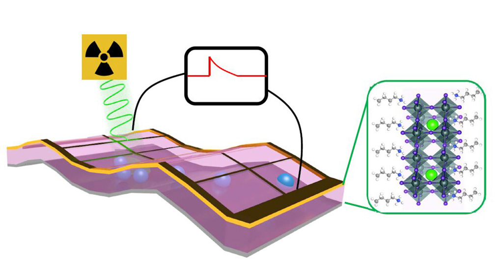
Image: Schematic of a perovskite thin film X-ray photon detector (Photo courtesy of Los Alamos National Laboratory)
A new generation of x-ray detectors based on a thin film of the mineral perovskite could transform future x-ray imaging technologies, according to a new study.
Developed at Los Alamos National Laboratory (LANL; NM, USA) and Argonne National Laboratory (ANL; Lemont, IL, USA), the thin-film x-ray detectors are comprised of highly crystalline, Ruddlesden-Popper phase layered perovskites that are over a hundred times more sensitive than conventional silicon-based detectors. Also, as the perovskite detectors are photocurrent devices, they do not require an outside power source to operate; instead, they work by generating substantial x-ray photon–induced open-circuit voltages in response to bombardment by X-ray beams.
The Perovskite films can be deposited on surfaces by spraying solutions that cure and leave thin layers of the material behind. As a result, the thin-layer detectors will be much easier and cheaper to produce than silicon-based detectors, which require high-temperature metal deposition under strict vacuum conditions. The lower-energy, increased-resolution detectors could also revolutionize security scanners and imaging in high-energy research facilities, for example those that use synchrotron light sources. The study was published on April 10, 2020, in Science Advances.
“The perovskite material at the heart of our detector prototype can be produced with low-cost fabrication techniques. Potentially, we could use ink-jet types of systems to print large scale detectors,” said lead author Hsinhan (Dave) Tsai, PhD, of the LANL department of materials physics and applications. “The result is a cost-effective, highly sensitive, and self-powered detector that could radically improve existing X-ray detectors, and potentially lead to a host of unforeseen applications.”
Perovskite, named after Russian mineralogist Lev Perovski (1792–1856), is a mineral that was discovered in the Ural Mountains by Gustav Rose in 1839. It is composed mainly of calcium titanate, and is rich in heavy elements, such as lead and iodine. As a result, X-rays that easily pass through silicon undetected are more readily absorbed, and detected, in perovskite.
Related Links:
Los Alamos National Laboratory
Argonne National Laboratory
Latest Radiography News
- AI Detects Early Signs of Aging from Chest X-Rays
- X-Ray Breakthrough Captures Three Image-Contrast Types in Single Shot
- AI Generates Future Knee X-Rays to Predict Osteoarthritis Progression Risk
- AI Algorithm Uses Mammograms to Accurately Predict Cardiovascular Risk in Women
- AI Hybrid Strategy Improves Mammogram Interpretation
- AI Technology Predicts Personalized Five-Year Risk of Developing Breast Cancer
- RSNA AI Challenge Models Can Independently Interpret Mammograms
- New Technique Combines X-Ray Imaging and Radar for Safer Cancer Diagnosis
- New AI Tool Helps Doctors Read Chest X‑Rays Better
- Wearable X-Ray Imaging Detecting Fabric to Provide On-The-Go Diagnostic Scanning
- AI Helps Radiologists Spot More Lesions in Mammograms
- AI Detects Fatty Liver Disease from Chest X-Rays
- AI Detects Hidden Heart Disease in Existing CT Chest Scans
- Ultra-Lightweight AI Model Runs Without GPU to Break Barriers in Lung Cancer Diagnosis
- AI Radiology Tool Identifies Life-Threatening Conditions in Milliseconds

- Machine Learning Algorithm Identifies Cardiovascular Risk from Routine Bone Density Scans
Channels
MRI
view channel
Novel Imaging Approach to Improve Treatment for Spinal Cord Injuries
Vascular dysfunction in the spinal cord contributes to multiple neurological conditions, including traumatic injuries and degenerative cervical myelopathy, where reduced blood flow can lead to progressive... Read more
AI-Assisted Model Enhances MRI Heart Scans
A cardiac MRI can reveal critical information about the heart’s function and any abnormalities, but traditional scans take 30 to 90 minutes and often suffer from poor image quality due to patient movement.... Read more
AI Model Outperforms Doctors at Identifying Patients Most At-Risk of Cardiac Arrest
Hypertrophic cardiomyopathy is one of the most common inherited heart conditions and a leading cause of sudden cardiac death in young individuals and athletes. While many patients live normal lives, some... Read moreUltrasound
view channel
Wearable Ultrasound Imaging System to Enable Real-Time Disease Monitoring
Chronic conditions such as hypertension and heart failure require close monitoring, yet today’s ultrasound imaging is largely confined to hospitals and short, episodic scans. This reactive model limits... Read more
Ultrasound Technique Visualizes Deep Blood Vessels in 3D Without Contrast Agents
Producing clear 3D images of deep blood vessels has long been difficult without relying on contrast agents, CT scans, or MRI. Standard ultrasound typically provides only 2D cross-sections, limiting clinicians’... Read moreNuclear Medicine
view channel
PET Imaging of Inflammation Predicts Recovery and Guides Therapy After Heart Attack
Acute myocardial infarction can trigger lasting heart damage, yet clinicians still lack reliable tools to identify which patients will regain function and which may develop heart failure.... Read more
Radiotheranostic Approach Detects, Kills and Reprograms Aggressive Cancers
Aggressive cancers such as osteosarcoma and glioblastoma often resist standard therapies, thrive in hostile tumor environments, and recur despite surgery, radiation, or chemotherapy. These tumors also... Read more
New Imaging Solution Improves Survival for Patients with Recurring Prostate Cancer
Detecting recurrent prostate cancer remains one of the most difficult challenges in oncology, as standard imaging methods such as bone scans and CT scans often fail to accurately locate small or early-stage tumors.... Read moreGeneral/Advanced Imaging
view channel
AI-Based Tool Accelerates Detection of Kidney Cancer
Diagnosing kidney cancer depends on computed tomography scans, often using contrast agents to reveal abnormalities in kidney structure. Tumors are not always searched for deliberately, as many scans are... Read more
New Algorithm Dramatically Speeds Up Stroke Detection Scans
When patients arrive at emergency rooms with stroke symptoms, clinicians must rapidly determine whether the cause is a blood clot or a brain bleed, as treatment decisions depend on this distinction.... Read moreImaging IT
view channel
New Google Cloud Medical Imaging Suite Makes Imaging Healthcare Data More Accessible
Medical imaging is a critical tool used to diagnose patients, and there are billions of medical images scanned globally each year. Imaging data accounts for about 90% of all healthcare data1 and, until... Read more
Global AI in Medical Diagnostics Market to Be Driven by Demand for Image Recognition in Radiology
The global artificial intelligence (AI) in medical diagnostics market is expanding with early disease detection being one of its key applications and image recognition becoming a compelling consumer proposition... Read moreIndustry News
view channel
GE HealthCare and NVIDIA Collaboration to Reimagine Diagnostic Imaging
GE HealthCare (Chicago, IL, USA) has entered into a collaboration with NVIDIA (Santa Clara, CA, USA), expanding the existing relationship between the two companies to focus on pioneering innovation in... Read more
Patient-Specific 3D-Printed Phantoms Transform CT Imaging
New research has highlighted how anatomically precise, patient-specific 3D-printed phantoms are proving to be scalable, cost-effective, and efficient tools in the development of new CT scan algorithms... Read more
Siemens and Sectra Collaborate on Enhancing Radiology Workflows
Siemens Healthineers (Forchheim, Germany) and Sectra (Linköping, Sweden) have entered into a collaboration aimed at enhancing radiologists' diagnostic capabilities and, in turn, improving patient care... Read more


