Artificial Intelligence (AI) Program Accurately Predicts Lung Cancer Risk from CT Images
By MedImaging International staff writers
Posted on 16 May 2021
An artificial intelligence (AI) program accurately predicts the risk that lung nodules detected on screening CT will become cancerous, according to a study published in the journal Radiology.Posted on 16 May 2021
In the new study, researchers at the Radboud University Medical Center (Nijmegen, The Netherlands) developed an algorithm for lung nodule assessment using deep learning, an AI application capable of finding certain patterns in imaging data. The researchers trained the algorithm on CT images of more than 16,000 nodules, including 1,249 malignancies, from the National Lung Screening Trial. They validated the algorithm on three large sets of imaging data of nodules from the Danish Lung Cancer Screening Trial.

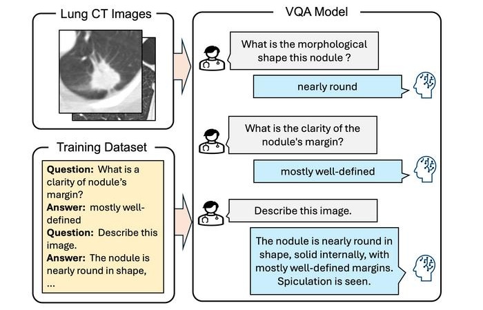
Illustration
The deep learning algorithm delivered excellent results, outperforming the established Pan-Canadian Early Detection of Lung Cancer model for lung nodule malignancy risk estimation. It performed comparably to 11 clinicians, including four thoracic radiologists, five radiology residents and two pulmonologists. The researchers plan to continue improving the algorithm by incorporating clinical parameters like age, sex and smoking history. They are also working on a deep learning algorithm that takes multiple CT examinations as input. The current algorithm is highly suitable for analyzing nodules at the initial, or baseline, screening, but for nodules detected at subsequent screenings, growth and appearance in comparison to the previous CT are important. The researchers have developed other algorithms to reliably extract imaging features from the chest CT related to chronic obstructive pulmonary diseases and cardiovascular diseases. They will be investigating how to effectively integrate these imaging features into the current algorithm.
“The algorithm may aid radiologists in accurately estimating the malignancy risk of pulmonary nodules,” said the study’s first author, Kiran Vaidhya Venkadesh, a Ph.D. candidate with the Diagnostic Image Analysis Group at Radboud University Medical Center in Nijmegen, the Netherlands. “This may help in optimizing follow-up recommendations for lung cancer screening participants.”
“As it does not require manual interpretation of nodule imaging characteristics, the proposed algorithm may reduce the substantial interobserver variability in CT interpretation,” said senior author Colin Jacobs, Ph.D., assistant professor in the Department of Medical Imaging at Radboud University Medical Center in Nijmegen. “This may lead to fewer unnecessary diagnostic interventions, lower radiologists’ workload and reduce costs of lung cancer screening.”
Related Links:
Radboud University Medical Center

Digital X-Ray Detector Panel
Acuity G4

X-ray Diagnostic System
FDX Visionary-A

Ultrasound Table
Women’s Ultrasound EA Table

Digital Intelligent Ferromagnetic Detector
Digital Ferromagnetic Detector