Fujifilm SonoSite Exhibits Complete Point-of-Care Ultrasound Portfolio at RSNA 2019
|
By MedImaging International staff writers Posted on 02 Dec 2019 |

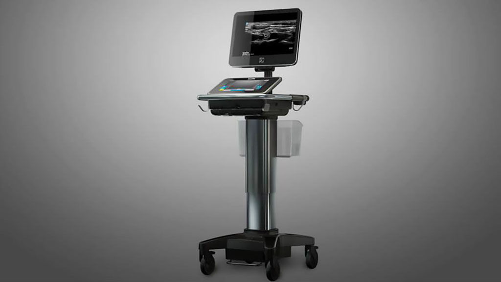
Image: SonoSite X-Porte (Photo courtesy of FUJIFILM SonoSite, Inc.)
FUJIFILM SonoSite, Inc. (Bothell, WA, USA), a developer of cutting-edge, point-of-care ultrasound (POCUS), exhibited numerous solutions at the annual meeting of the Radiological Society of North America (RSNA) held from December 1-5, 2019 in Chicago, USA.
FUJIFILM SonoSite’s portable, compact systems are expanding the use of ultrasound across the clinical spectrum by cost-effectively bringing high-performance ultrasound to the point of patient care. FUJIFILM SonoSite solutions that were demonstrated at RSNA 2019 included SonoSite X-Porte, a highly portable kiosk ultrasound system that fluidly combines striking image clarity with touchscreen controls and a customizable interface. It offers more than 80 real-time educational visual guides and tutorials. Proprietary high-definition imaging technology focuses the ultrasound beams with pinpoint precision, reducing artifact clutter and enhancing contrast resolution. FUJIFILM exhibited the SonoSite Edge II which offers an enhanced imaging experience through industry-first transducer innovations like Armored Cable Technology. In a clamshell design, it features an intuitive interface for easier access to frequently used functions and a wide-angle display with an anti-reflection coating for minimal adjustments during viewing. It is designed to be truly portable and used in the most rigorous environments.
Also featured at the event was SonoSite SII which empowers efficiency for clinicians through a simple portrait display, and smart user interface that adapts to the user’s imaging needs. FUJIFILM’s enhanced imaging technology on select transducers provides users with increased resolution and penetration, while maintaining durability and reliability with Armored Cable Technology. Other featured products included the SonoSite iViz, a powerful diagnostic tool that fits in the palm of the hand and provides quick answers in tough clinical environments, both at the bedside and in the field. It combines superior imaging performance, ultra-mobility, and one-handed operation while allowing collaboration and sharing of information with colleagues. FUJIFILM also demonstrated the SonoSite Synchronicity workflow manager that helps healthcare organizations optimize workflows, maximize financial return, improve quality assurance efficiency, and streamline credentialing processes. Built specifically for POCUS, SonoSite Synchronicity workflow manager securely centralizes exam data and standardizes clinical workflow while delivering administrative efficiencies. Additional features include built-in, customizable worksheets, intuitive dashboards, and the ability to access the tool from a computer, tablet or mobile device. Easily installed and scalable, SonoSite Synchronicity workflow manager was engineered to meet every organization’s unique requirements for standardization, consistency, and compliance across entire medical networks.
“In a healthcare world that’s increasingly complex, we help remove barriers so clinicians can concentrate on what really matters – patient care,” said Rich Fabian, President and Chief Operating Officer of FUJIFILM SonoSite, Inc. “Fujifilm SonoSite is not only committed to developing best in class ultrasound solutions, but also to enhancing education among clinicians who use POCUS all over the world.”
Related Links:
FUJIFILM SonoSite, Inc.
Latest RSNA 2019 News
- Carestream Introduces Three-Dimensional Extension of General Radiography Through Its Digital Tomosynthesis Functionality
- Lunit Demonstrates Latest Updated AI Solutions for Chest and Breast Radiology at RSNA 2019
- Bracco Diagnostics Unveils Contrast Media and Device Offerings at RSNA 2019
- Guerbet Showcases New Dose&Care and Other Digital Solutions with Diagnostic and Interventional Imaging Offerings
- Canon Introduces New Wireless Detectors and Digital PET/CT Scanner at RSNA 2019
- Siemens Healthineers Introduces SOMATOM On.site Mobile Head CT Scanner and AI-based MRI Assistants at RSNA
- Hologic Launches Unifi Workspace, Comprehensive Reading Solution for Breast Health Diagnostics
- Agfa Launches New Groundbreaking Digital Radiography Unit at RSNA 2019
- Fujifilm Previews World's First Glass-Free Digital Radiography Detector at RSNA 2019 Image
- NVIDIA Showcases Latest AI-driven Medical Imaging Advancements at RSNA 2019
- Philips Healthcare Demonstrates How AI Breast Software Brings Intelligence and Automation to Breast Ultrasound
- Siemens Healthineers Focuses on Digital Transformation of Imaging and Therapy at RSNA 2019
Channels
Radiography
view channel
Routine Mammograms Could Predict Future Cardiovascular Disease in Women
Mammograms are widely used to screen for breast cancer, but they may also contain overlooked clues about cardiovascular health. Calcium deposits in the arteries of the breast signal stiffening blood vessels,... Read more
AI Detects Early Signs of Aging from Chest X-Rays
Chronological age does not always reflect how fast the body is truly aging, and current biological age tests often rely on DNA-based markers that may miss early organ-level decline. Detecting subtle, age-related... Read moreMRI
view channel
MRI Scans Reveal Signature Patterns of Brain Activity to Predict Recovery from TBI
Recovery after traumatic brain injury (TBI) varies widely, with some patients regaining full function while others are left with lasting disabilities. Prognosis is especially difficult to assess in patients... Read more
Novel Imaging Approach to Improve Treatment for Spinal Cord Injuries
Vascular dysfunction in the spinal cord contributes to multiple neurological conditions, including traumatic injuries and degenerative cervical myelopathy, where reduced blood flow can lead to progressive... Read more
AI-Assisted Model Enhances MRI Heart Scans
A cardiac MRI can reveal critical information about the heart’s function and any abnormalities, but traditional scans take 30 to 90 minutes and often suffer from poor image quality due to patient movement.... Read more
AI Model Outperforms Doctors at Identifying Patients Most At-Risk of Cardiac Arrest
Hypertrophic cardiomyopathy is one of the most common inherited heart conditions and a leading cause of sudden cardiac death in young individuals and athletes. While many patients live normal lives, some... Read moreUltrasound
view channel
Imaging Technique Generates Simultaneous 3D Color Images of Soft-Tissue Structure and Vasculature
Medical imaging tools often force clinicians to choose between speed, structural detail, and functional insight. Ultrasound is fast and affordable but typically limited to two-dimensional anatomy, while... Read more
Wearable Ultrasound Imaging System to Enable Real-Time Disease Monitoring
Chronic conditions such as hypertension and heart failure require close monitoring, yet today’s ultrasound imaging is largely confined to hospitals and short, episodic scans. This reactive model limits... Read more
Ultrasound Technique Visualizes Deep Blood Vessels in 3D Without Contrast Agents
Producing clear 3D images of deep blood vessels has long been difficult without relying on contrast agents, CT scans, or MRI. Standard ultrasound typically provides only 2D cross-sections, limiting clinicians’... Read moreNuclear Medicine
view channel
Radiopharmaceutical Molecule Marker to Improve Choice of Bladder Cancer Therapies
Targeted cancer therapies only work when tumor cells express the specific molecular structures they are designed to attack. In urothelial carcinoma, a common form of bladder cancer, the cell surface protein... Read more
Cancer “Flashlight” Shows Who Can Benefit from Targeted Treatments
Targeted cancer therapies can be highly effective, but only when a patient’s tumor expresses the specific protein the treatment is designed to attack. Determining this usually requires biopsies or advanced... Read moreGeneral/Advanced Imaging
view channel
AI Tool Offers Prognosis for Patients with Head and Neck Cancer
Oropharyngeal cancer is a form of head and neck cancer that can spread through lymph nodes, significantly affecting survival and treatment decisions. Current therapies often involve combinations of surgery,... Read more
New 3D Imaging System Addresses MRI, CT and Ultrasound Limitations
Medical imaging is central to diagnosing and managing injuries, cancer, infections, and chronic diseases, yet existing tools each come with trade-offs. Ultrasound, X-ray, CT, and MRI can be costly, time-consuming,... Read moreImaging IT
view channel
New Google Cloud Medical Imaging Suite Makes Imaging Healthcare Data More Accessible
Medical imaging is a critical tool used to diagnose patients, and there are billions of medical images scanned globally each year. Imaging data accounts for about 90% of all healthcare data1 and, until... Read more
Global AI in Medical Diagnostics Market to Be Driven by Demand for Image Recognition in Radiology
The global artificial intelligence (AI) in medical diagnostics market is expanding with early disease detection being one of its key applications and image recognition becoming a compelling consumer proposition... Read moreIndustry News
view channel
GE HealthCare and NVIDIA Collaboration to Reimagine Diagnostic Imaging
GE HealthCare (Chicago, IL, USA) has entered into a collaboration with NVIDIA (Santa Clara, CA, USA), expanding the existing relationship between the two companies to focus on pioneering innovation in... Read more
Patient-Specific 3D-Printed Phantoms Transform CT Imaging
New research has highlighted how anatomically precise, patient-specific 3D-printed phantoms are proving to be scalable, cost-effective, and efficient tools in the development of new CT scan algorithms... Read more
Siemens and Sectra Collaborate on Enhancing Radiology Workflows
Siemens Healthineers (Forchheim, Germany) and Sectra (Linköping, Sweden) have entered into a collaboration aimed at enhancing radiologists' diagnostic capabilities and, in turn, improving patient care... Read more


