New AI Platform to Analyze NHS Radiological Scans
|
By MedImaging International staff writers Posted on 30 May 2019 |


|

Image: The DGX-2 two petaFLOPS supercomputer system (Photo courtesy of Nvidia).
A novel artificial intelligence (AI) system intended for the United Kingdom National Health Service (NHS; London) will automate nationwide radiological interpretation across multiple clinical pathways, including oncology, cardiology, and neurology.
The AI project is a joint effort of Nvidia (Santa Clara, CA, USA) and King’s College London (KCL; United Kingdom), which will build and train the AI platform to interpret radiological scans for hospitals across the UK. The technology would thus free up overworked specialists and could also lead to breakthroughs across the medical imaging landscape, from determining the root cause of various cancers to helping classify specific neurological impairments and identifying optimal treatment plans.
At the core of the first stage of the project is a graphics processing unit (GPU)-powered two-petaflops Nvidia DGX-2 supercomputer, combining 16 interconnected GPUs, which Nvidia claims is currently the world's most powerful AI system. The project will also employ the Nvidia Clara AI toolkit, an open-source NiftyNet image-analysis neural-network, and a host of supplemental imaging technologies from existing NHS partners such as Kheiron Medical (London, UK), Mirada (London, UK) and Scan.
“Together with King's College London, we're working to push the envelope in AI for healthcare,” said Jaap Zuiderveld, vice president for Europe, the Middle East and Africa (EMEA) at Nvidia. “DGX-2 systems with the Nvidia Clara platform will enable the project to scale and drive breakthroughs in radiology [and] ultimately help improve patient outcomes within the NHS.”
“This center marks a significant chapter in the future of AI-enabled NHS hospitals, and the infrastructure is an essential part of building new AI tools which will benefit patients and the healthcare system as a whole,” said Professor Sebastien Ourselin, PhD, head of the School of Biomedical Engineering & Imaging Sciences at KCL. “The Nvidia DGX-2 AI system's large memory and massive computing power make it possible for us to tackle training of large, 3D datasets in minutes instead of days while keeping the data secure on the premises of the hospital.”
As security and governance of data in clinical environments is of the highest importance, and since AI models within the project will be built from patient data from across the entire UK-wide NHS system, the center has decided to employ federated learning, which keeps data within its own, secure domain, while allowing algorithms to be developed at multiple sites using data located at hospitals around the UK. The federated learning approach is designed to ensure a robust and secure system that more than complies with government data-privacy requirements.




The AI project is a joint effort of Nvidia (Santa Clara, CA, USA) and King’s College London (KCL; United Kingdom), which will build and train the AI platform to interpret radiological scans for hospitals across the UK. The technology would thus free up overworked specialists and could also lead to breakthroughs across the medical imaging landscape, from determining the root cause of various cancers to helping classify specific neurological impairments and identifying optimal treatment plans.
At the core of the first stage of the project is a graphics processing unit (GPU)-powered two-petaflops Nvidia DGX-2 supercomputer, combining 16 interconnected GPUs, which Nvidia claims is currently the world's most powerful AI system. The project will also employ the Nvidia Clara AI toolkit, an open-source NiftyNet image-analysis neural-network, and a host of supplemental imaging technologies from existing NHS partners such as Kheiron Medical (London, UK), Mirada (London, UK) and Scan.
“Together with King's College London, we're working to push the envelope in AI for healthcare,” said Jaap Zuiderveld, vice president for Europe, the Middle East and Africa (EMEA) at Nvidia. “DGX-2 systems with the Nvidia Clara platform will enable the project to scale and drive breakthroughs in radiology [and] ultimately help improve patient outcomes within the NHS.”
“This center marks a significant chapter in the future of AI-enabled NHS hospitals, and the infrastructure is an essential part of building new AI tools which will benefit patients and the healthcare system as a whole,” said Professor Sebastien Ourselin, PhD, head of the School of Biomedical Engineering & Imaging Sciences at KCL. “The Nvidia DGX-2 AI system's large memory and massive computing power make it possible for us to tackle training of large, 3D datasets in minutes instead of days while keeping the data secure on the premises of the hospital.”
As security and governance of data in clinical environments is of the highest importance, and since AI models within the project will be built from patient data from across the entire UK-wide NHS system, the center has decided to employ federated learning, which keeps data within its own, secure domain, while allowing algorithms to be developed at multiple sites using data located at hospitals around the UK. The federated learning approach is designed to ensure a robust and secure system that more than complies with government data-privacy requirements.

Silver Member
X-Ray QA Device
Accu-Gold+ Touch Pro
Mobile X-Ray System
K4W
Digital Radiography System
DR-300
Medical Radiographic X-Ray Machine
TR30N HF
Latest Imaging IT News
- Interactive AI Tool Supports Explainable Lung Nodule Assessment
- Breast Imaging Software Enhances Visualization and Tissue Characterization in Challenging Cases
- New Google Cloud Medical Imaging Suite Makes Imaging Healthcare Data More Accessible
- Global AI in Medical Diagnostics Market to Be Driven by Demand for Image Recognition in Radiology
- AI-Based Mammography Triage Software Helps Dramatically Improve Interpretation Process
- Artificial Intelligence (AI) Program Accurately Predicts Lung Cancer Risk from CT Images
- Image Management Platform Streamlines Treatment Plans
- AI-Based Technology for Ultrasound Image Analysis Receives FDA Approval
- AI Technology for Detecting Breast Cancer Receives CE Mark Approval
- Digital Pathology Software Improves Workflow Efficiency
Channels
Radiography
view channel
Simple Chest X-Ray Measure Predicts Survival After Lung Cancer Surgery
Obstructive ventilatory disorder, marked by airflow limitation that reduces breathing efficiency, increases postoperative risk in patients with lung cancer. Although surgery offers the best chance of cure,... Read more
AI Detection Tool Improves Identification of Lobular Breast Cancer
Breast cancer screening seeks early detection, yet some subtypes remain difficult to visualize on mammography, risking delayed diagnosis. On average, 1 in 20 women worldwide will develop breast cancer,... Read more
MRI
view channel
Cardiac MRI Measure Improves Risk Prediction in Tricuspid Regurgitation
Tricuspid regurgitation, in which blood flows back from the right ventricle into the right atrium, can lead to progressive right-sided heart failure. Clinicians need reliable ways to gauge severity and... Read more
AI System Improves Accuracy of Cardiac MRI Interpretation
Cardiac magnetic resonance imaging (MRI) is the reference standard for assessing cardiac structure, function, and tissue health. Yet each exam can contain hundreds to thousands of images across multiple... Read more
Ultrasound
view channel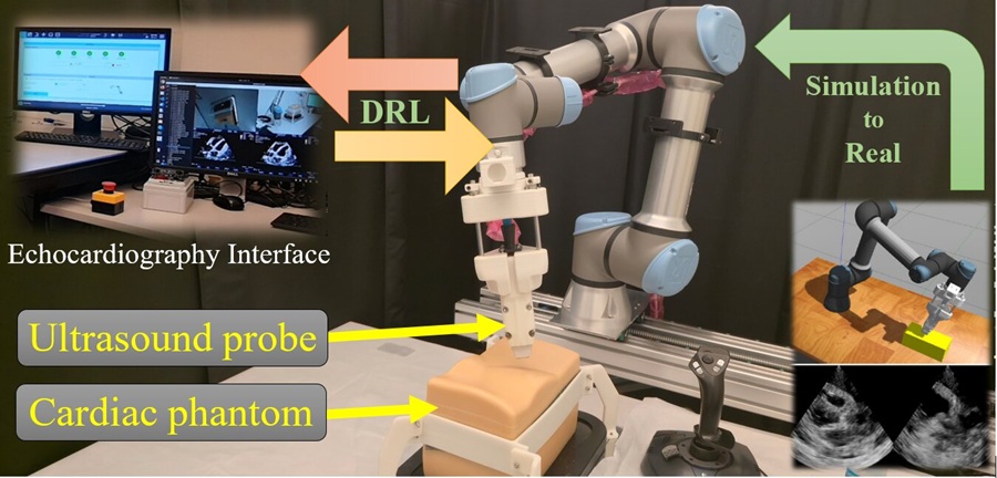
AI Robotic Ultrasound System Automates Echocardiography and Improves Consistency
Echocardiography, an ultrasound examination of the heart, is central to diagnosing and managing cardiovascular disease. Many services struggle with limited availability of skilled sonographers, variable... Read more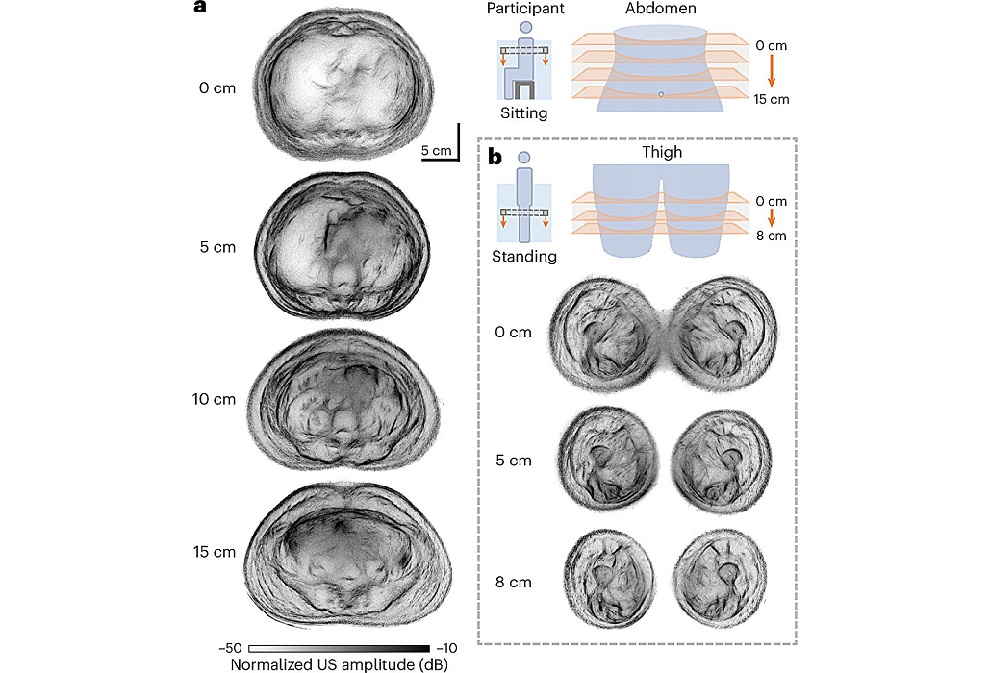
Whole Cross-Section Ultrasound System Enables Operator-Independent Imaging
Conventional ultrasound is central to bedside imaging but is limited by a narrow field of view and operator variability. Comprehensive cross-sectional assessment typically requires computed tomography... Read more
Nuclear Medicine
view channel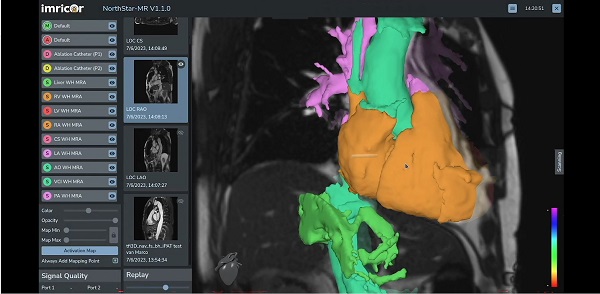
MR-Guided Cardiac Mapping System Enables Radiation-Free Procedures
Cardiac electrophysiology procedures are typically guided by X-ray fluoroscopy, which limits soft-tissue visualization and exposes patients and clinical staff to ionizing radiation. Real-time mapping that... Read more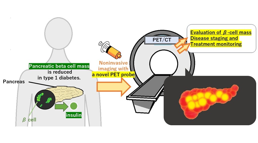
PET Tracer Enables Noninvasive Measurement of Beta Cell Mass
Type 1 diabetes is an autoimmune disease in which the immune system destroys insulin-producing pancreatic beta cells. Loss of these cells destabilizes glucose control and drives complications.... Read more
New Imaging Tool Sheds Light on Tumor Fat Metabolism
Rapidly growing tumors reprogram metabolism to meet high energy demands. While many cancers preferentially consume glucose, lipid utilization by malignant cells is difficult to measure in living subjects.... Read more
Radiopharmaceutical Molecule Marker to Improve Choice of Bladder Cancer Therapies
Targeted cancer therapies only work when tumor cells express the specific molecular structures they are designed to attack. In urothelial carcinoma, a common form of bladder cancer, the cell surface protein... Read moreGeneral/Advanced Imaging
view channel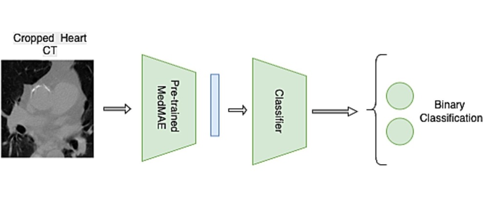
Multimodal AI Tool Combines CT and Health Records to Predict Heart Risk
Cardiovascular disease is a leading cause of death and an underrecognized risk for people treated for breast cancer. Cardiac complications can affect survival and quality of life. Clinicians need tools... Read more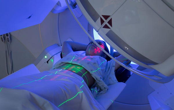
AI Tool Automates Radiotherapy Planning for Cervical and Prostate Cancer
Cervical cancer causes most of its global mortality in low- and middle-income countries, where radiotherapy capacity and specialist staff are limited. Treatment planning is labor-intensive and can delay... Read more
Imaging IT
view channel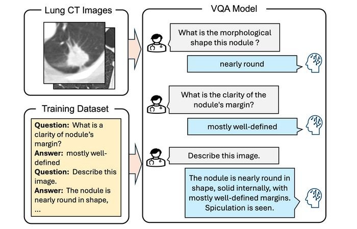
Interactive AI Tool Supports Explainable Lung Nodule Assessment
Lung cancer is a leading cause of cancer mortality, and timely characterization of pulmonary nodules on chest computed tomography (CT) is essential for directing care. Interpreting nodule morphology demands... Read more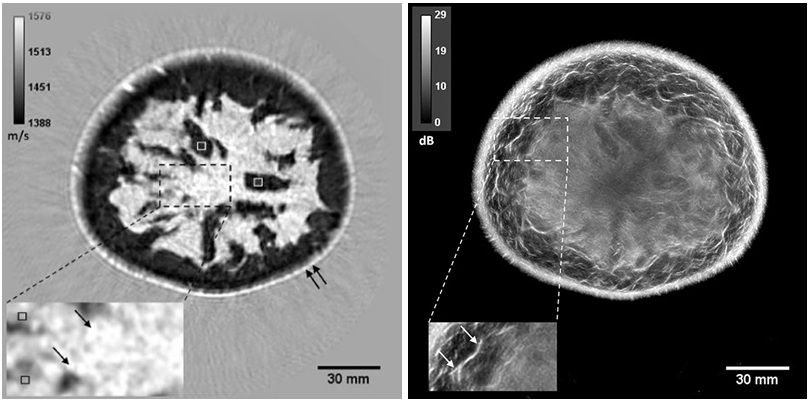
Breast Imaging Software Enhances Visualization and Tissue Characterization in Challenging Cases
Breast imaging can be particularly challenging in cases involving small breasts or implants, where image reconstruction and tissue characterization may be limited. Clinicians also need reproducible analysis... Read more
New Google Cloud Medical Imaging Suite Makes Imaging Healthcare Data More Accessible
Medical imaging is a critical tool used to diagnose patients, and there are billions of medical images scanned globally each year. Imaging data accounts for about 90% of all healthcare data1 and, until... Read more
Global AI in Medical Diagnostics Market to Be Driven by Demand for Image Recognition in Radiology
The global artificial intelligence (AI) in medical diagnostics market is expanding with early disease detection being one of its key applications and image recognition becoming a compelling consumer proposition... Read moreIndustry News
view channel
GE HealthCare Highlights AI-Supported Radiation Therapy Tools at ESTRO 2026
At the European Society for Radiotherapy and Oncology (ESTRO) 2026 Congress in Stockholm, GE HealthCare is highlighting Intelligent Radiation Therapy (iRT), MIM Software innovations, and BK Medical surgical... Read more