Diagnostic Image Display Designed for Both PACS and Breast Imaging
|
By MedImaging International staff writers Posted on 30 Oct 2014 |
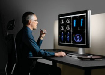
The first diagnostic display designed for both picture archiving and communication systems (PACS) and breast imaging provides excellent image quality, inventive productivity features, and a focus on ergonomics.
Barco’s (Kortrijk, Belgium) Coronis Uniti image display was devised to tackle specific challenges in modern radiology: increasing image volumes, growing complexity, and ergonomic stress. “With nearly 30 years of experience in healthcare imaging, we understand the radiology market and the challenges the industry is facing today. It’s this experience that led to the development of Coronis Uniti, a diagnostic display system that unifies the workflow for radiologists,” commented Lynda Domogalla, vice president of product marketing for Barco’s healthcare division.
Radiologists’ most significant issues were brought to light recently, when the healthcare research agency the MarkeTech Group (Davis CA, USA), a global marketing research company, revealed the findings of its most recent radiology survey. The study, which interviewed over 200 radiologists from Europe and North America, also offered distinct insights into possible solutions. Most of the respondents, for instance, indicated that higher image quality, a more efficient workflow, and increased comfort would significantly increase reading performance.
The features of the new Coronis Uniti display provides enhanced reading performance. By supporting both PACS and breast imaging, calibrated color and grayscale, two dimensions (2D) and 3D, and static as well as dynamic images, the display eliminates the need for multi-head display setups or moving to another workstation, thereby increasing workflow efficiency while cutting costs. Focused on driving productivity, Coronis Uniti is loaded with clever features that improve the reading experience. These include RapidFrame technology to provide clear and in-focus moving images and a novel multi-touch pad for fast application control.
The 12 MP color screen offers an extremely high resolution. Furthermore, to meet the DICOM standard for grayscale images and to guarantee consistent color, the display is equipped with Barco’s industry-changing SteadyColor calibration technology. Additional special features such as Color Per Pixel Uniformity to reduce screen noise and Ambient Light Compensation ensure that image quality is excellent, in any lighting environment. The MediCal QAWeb software for automated calibration and quality assurance (QA) assures enhanced performance without interruptions.
The display optimizes the reading experience by mirroring a human’s natural field of vision. Its carefully designed format minimizes the need for head and eye movements, while also creating the perfect canvas for side-by-side comparison of multiple images.
To reduce eye fatigue, the SoftGlow wall light adds ambient light to the reading room, while the SoftGlow task light shines a light on film folders and papers. Barco’s Optical Glass technology is also integrated into the display, which reduces reflections and enhances image sharpness for greater viewing comfort. Moreover, Coronis Uniti can be turned into a virtual lightbox to compare film-based prior studies—using a handy film clip—with current digital exams.
“With advances in image quality, productivity, and ergonomics, Barco is convinced that the Coronis Uniti display system will change the radiology reading room,” said Ms. Domogalla. “By simplifying and standardizing the image reading technology platform, it becomes easier to manage and control the entire display network. Furthermore, by replacing the multiple-head display setup, Coronis Uniti offers unique opportunities to reduce display cost, real estate, and operational expenditure. In short, this is a one-time investment that will last a lifetime.”
Barco, a global technology company, designs and develops visualization products for a range of selected professional markets.
Related Links:
Barco
MarkeTech Group
Read the full article by registering today, it's FREE! 

Register now for FREE to MedImaging.net and get access to news and events that shape the world of Radiology. 
- Free digital version edition of Medical Imaging International sent by email on regular basis
- Free print version of Medical Imaging International magazine (available only outside USA and Canada).
- Free and unlimited access to back issues of Medical Imaging International in digital format
- Free Medical Imaging International Newsletter sent every week containing the latest news
- Free breaking news sent via email
- Free access to Events Calendar
- Free access to LinkXpress new product services
- REGISTRATION IS FREE AND EASY!
Sign in: Registered website members
Sign in: Registered magazine subscribers
Latest Imaging IT News
- New Google Cloud Medical Imaging Suite Makes Imaging Healthcare Data More Accessible
- Global AI in Medical Diagnostics Market to Be Driven by Demand for Image Recognition in Radiology
- AI-Based Mammography Triage Software Helps Dramatically Improve Interpretation Process
- Artificial Intelligence (AI) Program Accurately Predicts Lung Cancer Risk from CT Images
- Image Management Platform Streamlines Treatment Plans
- AI-Based Technology for Ultrasound Image Analysis Receives FDA Approval
- AI Technology for Detecting Breast Cancer Receives CE Mark Approval
- Digital Pathology Software Improves Workflow Efficiency
- Patient-Centric Portal Facilitates Direct Imaging Access
- New Workstation Supports Customer-Driven Imaging Workflow
Channels
Radiography
view channel
Machine Learning Algorithm Identifies Cardiovascular Risk from Routine Bone Density Scans
A new study published in the Journal of Bone and Mineral Research reveals that an automated machine learning program can predict the risk of cardiovascular events and falls or fractures by analyzing bone... Read more
AI Improves Early Detection of Interval Breast Cancers
Interval breast cancers, which occur between routine screenings, are easier to treat when detected earlier. Early detection can reduce the need for aggressive treatments and improve the chances of better outcomes.... Read more
World's Largest Class Single Crystal Diamond Radiation Detector Opens New Possibilities for Diagnostic Imaging
Diamonds possess ideal physical properties for radiation detection, such as exceptional thermal and chemical stability along with a quick response time. Made of carbon with an atomic number of six, diamonds... Read moreMRI
view channel
New MRI Technique Reveals Hidden Heart Issues
Traditional exercise stress tests conducted within an MRI machine require patients to lie flat, a position that artificially improves heart function by increasing stroke volume due to gravity-driven blood... Read more
Shorter MRI Exam Effectively Detects Cancer in Dense Breasts
Women with extremely dense breasts face a higher risk of missed breast cancer diagnoses, as dense glandular and fibrous tissue can obscure tumors on mammograms. While breast MRI is recommended for supplemental... Read moreUltrasound
view channel
New Incision-Free Technique Halts Growth of Debilitating Brain Lesions
Cerebral cavernous malformations (CCMs), also known as cavernomas, are abnormal clusters of blood vessels that can grow in the brain, spinal cord, or other parts of the body. While most cases remain asymptomatic,... Read more.jpeg)
AI-Powered Lung Ultrasound Outperforms Human Experts in Tuberculosis Diagnosis
Despite global declines in tuberculosis (TB) rates in previous years, the incidence of TB rose by 4.6% from 2020 to 2023. Early screening and rapid diagnosis are essential elements of the World Health... Read moreNuclear Medicine
view channel
New Imaging Approach Could Reduce Need for Biopsies to Monitor Prostate Cancer
Prostate cancer is the second leading cause of cancer-related death among men in the United States. However, the majority of older men diagnosed with prostate cancer have slow-growing, low-risk forms of... Read more
Novel Radiolabeled Antibody Improves Diagnosis and Treatment of Solid Tumors
Interleukin-13 receptor α-2 (IL13Rα2) is a cell surface receptor commonly found in solid tumors such as glioblastoma, melanoma, and breast cancer. It is minimally expressed in normal tissues, making it... Read moreGeneral/Advanced Imaging
view channel
First-Of-Its-Kind Wearable Device Offers Revolutionary Alternative to CT Scans
Currently, patients with conditions such as heart failure, pneumonia, or respiratory distress often require multiple imaging procedures that are intermittent, disruptive, and involve high levels of radiation.... Read more
AI-Based CT Scan Analysis Predicts Early-Stage Kidney Damage Due to Cancer Treatments
Radioligand therapy, a form of targeted nuclear medicine, has recently gained attention for its potential in treating specific types of tumors. However, one of the potential side effects of this therapy... Read moreIndustry News
view channel
GE HealthCare and NVIDIA Collaboration to Reimagine Diagnostic Imaging
GE HealthCare (Chicago, IL, USA) has entered into a collaboration with NVIDIA (Santa Clara, CA, USA), expanding the existing relationship between the two companies to focus on pioneering innovation in... Read more
Patient-Specific 3D-Printed Phantoms Transform CT Imaging
New research has highlighted how anatomically precise, patient-specific 3D-printed phantoms are proving to be scalable, cost-effective, and efficient tools in the development of new CT scan algorithms... Read more
Siemens and Sectra Collaborate on Enhancing Radiology Workflows
Siemens Healthineers (Forchheim, Germany) and Sectra (Linköping, Sweden) have entered into a collaboration aimed at enhancing radiologists' diagnostic capabilities and, in turn, improving patient care... Read more


