New Software Designed to Ensure Faster Care, Treatment for Stroke Patients
|
By MedImaging International staff writers Posted on 29 Apr 2014 |
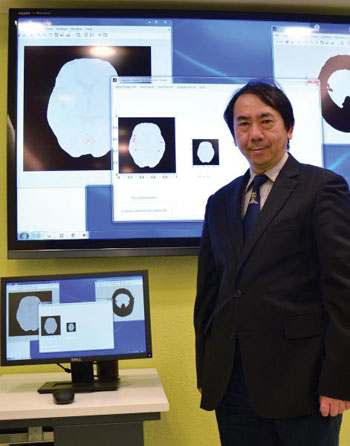
Image: Dr. Fuk-hay Tang believes that the CAD stroke system raises hope for patients with ischemic stroke by detecting signs of stroke from computed tomography (CT) scans (Photo courtesy of the Hong Kong Polytechnic University).
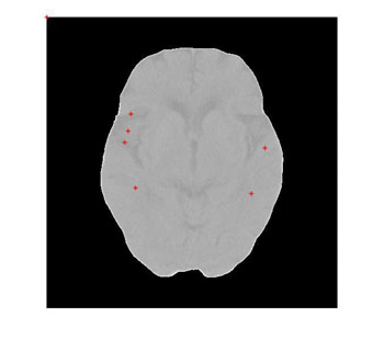
Image: Automatic detection of potential ischemic stroke areas (Photo courtesy of the Hong Kong Polytechnic University).
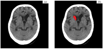
Image: CT scan without CAD (a) CT scan with CAD result (b) (Photo courtesy of the Hong Kong Polytechnic University).
New computer technology has the potential to treat people affected by ischemic stroke. Developed by researchers in Hong Kong, this innovative application that proficiently analyses brain scans could save lives by helping clinicians determine if a patient has the life-threatening disorder.
The computer-aided design (CAD) stroke technology is capable of detecting signs of stroke from computed tomography (CT) scans. When blood flow to the brain is blocked, an area of the brain turns softer or decreases in density due to insufficient blood flow, pointing to an ischemic stroke, which is a very common kind of stroke that accounts for over 80% of overall stroke cases.
As demonstrated by Dr. Fuk-hay Tang from the department of health technology and informatics at Hong Kong Polytechnic University (PolyU), CT scans are fed into the CAD stroke computer, which will make calculations and comparisons to locate regions suspected of insufficient blood flow. In 10 minutes, scans with highlighted areas of abnormality will come out for a physician’s review. Early alteration including loss of insular ribbon, loss of sulcus and dense MCA signs can be identified, helping clinicians determine if blood clots are present.
A diagnostic tool that can accelerate the process will be greatly helpful in saving lives. Dr. Tang stated, “The clock is ticking for stroke patients. Medications taken in three hours from the onset of stroke are deemed most effective. Chances of recovery decrease with every minute passing by. It typically takes 30 minutes at best for the ambulance to arrive at the hospital. Then, another 45 minutes to one hour are then required for CT or magnetic resonance imaging [MRI] scans after the patient has been checked and dispatched for the test, which means some waiting, and time will slip by. Afterwards, the brain scan will take another 10 to 15 minutes. If our tool can help doctors arrive at a diagnosis in 10 minutes, the shorter response time will make meeting the target more achievable. It might come in handy for physicians with less experience in stroke, and patient care can be maintained in hospitals where human and other vital resources are already stretched to the limit.”
The life-saving tool can also identify slight and tiny changes in the brain that would escape the eye of even an experienced specialist, slashing the chances of missed diagnosis. False-positive and false-negative cases, and other less serious disorders that mimic a stroke can also be ruled out, allowing a fully-informed decision to be made.
Furthermore, equipped with the built-in artificial intelligence feature, the CAD stroke technology would learn by experience. With every scan passing through, along with feedback from stroke specialists, the application will improve on its accuracy over time. “It is important to identify stroke patients and help them get the urgent treatment they need,” said Dr .Tang. “Prompt and accurate diagnosis is in the forefront of our minds when designing the medical application. Healthcare professionals should focus on what they do best and let us take care of the rest.”
Related Links:
Hong Kong Polytechnic University
Latest Imaging IT News
- New Google Cloud Medical Imaging Suite Makes Imaging Healthcare Data More Accessible
- Global AI in Medical Diagnostics Market to Be Driven by Demand for Image Recognition in Radiology
- AI-Based Mammography Triage Software Helps Dramatically Improve Interpretation Process
- Artificial Intelligence (AI) Program Accurately Predicts Lung Cancer Risk from CT Images
- Image Management Platform Streamlines Treatment Plans
- AI-Based Technology for Ultrasound Image Analysis Receives FDA Approval
- AI Technology for Detecting Breast Cancer Receives CE Mark Approval
- Digital Pathology Software Improves Workflow Efficiency
- Patient-Centric Portal Facilitates Direct Imaging Access
- New Workstation Supports Customer-Driven Imaging Workflow
Channels
Radiography
view channel
Machine Learning Algorithm Identifies Cardiovascular Risk from Routine Bone Density Scans
A new study published in the Journal of Bone and Mineral Research reveals that an automated machine learning program can predict the risk of cardiovascular events and falls or fractures by analyzing bone... Read more
AI Improves Early Detection of Interval Breast Cancers
Interval breast cancers, which occur between routine screenings, are easier to treat when detected earlier. Early detection can reduce the need for aggressive treatments and improve the chances of better outcomes.... Read more
World's Largest Class Single Crystal Diamond Radiation Detector Opens New Possibilities for Diagnostic Imaging
Diamonds possess ideal physical properties for radiation detection, such as exceptional thermal and chemical stability along with a quick response time. Made of carbon with an atomic number of six, diamonds... Read moreMRI
view channel
New MRI Technique Reveals Hidden Heart Issues
Traditional exercise stress tests conducted within an MRI machine require patients to lie flat, a position that artificially improves heart function by increasing stroke volume due to gravity-driven blood... Read more
Shorter MRI Exam Effectively Detects Cancer in Dense Breasts
Women with extremely dense breasts face a higher risk of missed breast cancer diagnoses, as dense glandular and fibrous tissue can obscure tumors on mammograms. While breast MRI is recommended for supplemental... Read moreUltrasound
view channel
New Incision-Free Technique Halts Growth of Debilitating Brain Lesions
Cerebral cavernous malformations (CCMs), also known as cavernomas, are abnormal clusters of blood vessels that can grow in the brain, spinal cord, or other parts of the body. While most cases remain asymptomatic,... Read more.jpeg)
AI-Powered Lung Ultrasound Outperforms Human Experts in Tuberculosis Diagnosis
Despite global declines in tuberculosis (TB) rates in previous years, the incidence of TB rose by 4.6% from 2020 to 2023. Early screening and rapid diagnosis are essential elements of the World Health... Read moreNuclear Medicine
view channel
New Imaging Approach Could Reduce Need for Biopsies to Monitor Prostate Cancer
Prostate cancer is the second leading cause of cancer-related death among men in the United States. However, the majority of older men diagnosed with prostate cancer have slow-growing, low-risk forms of... Read more
Novel Radiolabeled Antibody Improves Diagnosis and Treatment of Solid Tumors
Interleukin-13 receptor α-2 (IL13Rα2) is a cell surface receptor commonly found in solid tumors such as glioblastoma, melanoma, and breast cancer. It is minimally expressed in normal tissues, making it... Read moreGeneral/Advanced Imaging
view channel
CT Colonography Beats Stool DNA Testing for Colon Cancer Screening
As colorectal cancer remains the second leading cause of cancer-related deaths worldwide, early detection through screening is vital to reduce advanced-stage treatments and associated costs.... Read more
First-Of-Its-Kind Wearable Device Offers Revolutionary Alternative to CT Scans
Currently, patients with conditions such as heart failure, pneumonia, or respiratory distress often require multiple imaging procedures that are intermittent, disruptive, and involve high levels of radiation.... Read more
AI-Based CT Scan Analysis Predicts Early-Stage Kidney Damage Due to Cancer Treatments
Radioligand therapy, a form of targeted nuclear medicine, has recently gained attention for its potential in treating specific types of tumors. However, one of the potential side effects of this therapy... Read moreIndustry News
view channel
GE HealthCare and NVIDIA Collaboration to Reimagine Diagnostic Imaging
GE HealthCare (Chicago, IL, USA) has entered into a collaboration with NVIDIA (Santa Clara, CA, USA), expanding the existing relationship between the two companies to focus on pioneering innovation in... Read more
Patient-Specific 3D-Printed Phantoms Transform CT Imaging
New research has highlighted how anatomically precise, patient-specific 3D-printed phantoms are proving to be scalable, cost-effective, and efficient tools in the development of new CT scan algorithms... Read more
Siemens and Sectra Collaborate on Enhancing Radiology Workflows
Siemens Healthineers (Forchheim, Germany) and Sectra (Linköping, Sweden) have entered into a collaboration aimed at enhancing radiologists' diagnostic capabilities and, in turn, improving patient care... Read more


