Researchers Use PET to Visualize Cardiac Amyloidosis
|
By Andrew Deutsch Posted on 22 Nov 2016 |

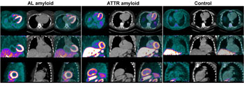
Image: Researchers have demonstrated that F-18-florbetaben PET imaging can be used to accurately differentiate between cardiac amyloidosis and hypertensive heart disease (Photo courtesy of W. Phillip Law/Princess Alexandra Hospital, Brisbane, Australia).
Results of a study have shown that a new non-invasive technique using PET imaging and a radiotracer, can visualize abnormal protein deposits in the heart, in a condition called cardiac amyloidosis.
The researchers used fluorine-18 (F-18)-florbetaben, a radioactive tracer, and Positron Emission Tomography (PET) to visualize and quantify the deposition of amyloid proteins in heart muscle. The technique could be used to diagnose cardiac amyloidosis accurately and non-invasively, and could also help clinicians the monitor disease burden.
The study results were published in the November 2016 issue of The Journal of Nuclear Medicine. The researchers from the Princess Alexandra Hospital (Brisbane, Australia) performed F-18-florbetaben PET in 14 subjects and compared patients who had thickened heart muscle, secondary to amyloid deposition, with those that had thickened myocardium as a result of hypertensive heart disease.
The researchers found a higher target-to-background Standardized Uptake Values (SUV) ratio and percentage myocardial radiotracer retention in amyloid patients compared to the control subjects with heart disease. The results show that the new PET technique can be used to identify and differentiate between patients with cardiac amyloidosis and those with hypertensive heart disease.
Corresponding author of the study, Dr. W. Phillip Law, said, "The first signs and symptoms of the disease are non-specific and usually attributed to other conditions. Currently, there is no definitive test to diagnose cardiac amyloidosis other than an invasive biopsy of the heart muscle. Cardiac amyloidosis is often not diagnosed until late in the course of the disease, as the typical appearance of the infiltrated myocardium on echocardiography and MRI can be mistaken for other more prevalent disorders. Tailored molecular imaging with PET using florbetaben may significantly simplify the diagnostic algorithm for patients with suspected cardiac amyloidosis. Future studies investigating florbetaben uptake pattern in other [non-amyloid, non-hypertensive] causes of heart muscle thickening would further clarify the specificity of florbetaben. The relationship of PET quantification of florbetaben retention in the heart, with histological amyloid plaque burden, may provide another means of monitoring disease and could also be useful in monitoring response of cardiac amyloid to treatment, but further research needs to be undertaken to investigate this relationship."
Related Links:
Princess Alexandra Hospital
Latest Nuclear Medicine News
- New PET Biomarker Predicts Success of Immune Checkpoint Blockade Therapy
- New PET Agent Rapidly and Accurately Visualizes Lesions in Clear Cell Renal Cell Carcinoma Patients
- New Imaging Technique Monitors Inflammation Disorders without Radiation Exposure
- New SPECT/CT Technique Could Change Imaging Practices and Increase Patient Access
- New Radiotheranostic System Detects and Treats Ovarian Cancer Noninvasively
- AI System Automatically and Reliably Detects Cardiac Amyloidosis Using Scintigraphy Imaging
- Early 30-Minute Dynamic FDG-PET Acquisition Could Halve Lung Scan Times
- New Method for Triggering and Imaging Seizures to Help Guide Epilepsy Surgery
- Radioguided Surgery Accurately Detects and Removes Metastatic Lymph Nodes in Prostate Cancer Patients
- New PET Tracer Detects Inflammatory Arthritis Before Symptoms Appear
- Novel PET Tracer Enhances Lesion Detection in Medullary Thyroid Cancer
- Targeted Therapy Delivers Radiation Directly To Cells in Hard-To-Treat Cancers
- New PET Tracer Noninvasively Identifies Cancer Gene Mutation for More Precise Diagnosis
- Algorithm Predicts Prostate Cancer Recurrence in Patients Treated by Radiation Therapy
- Novel PET Imaging Tracer Noninvasively Identifies Cancer Gene Mutation for More Precise Diagnosis
- Ultrafast Laser Technology to Improve Cancer Treatment
Channels
Radiography
view channel
Novel Breast Imaging System Proves As Effective As Mammography
Breast cancer remains the most frequently diagnosed cancer among women. It is projected that one in eight women will be diagnosed with breast cancer during her lifetime, and one in 42 women who turn 50... Read more
AI Assistance Improves Breast-Cancer Screening by Reducing False Positives
Radiologists typically detect one case of cancer for every 200 mammograms reviewed. However, these evaluations often result in false positives, leading to unnecessary patient recalls for additional testing,... Read moreMRI
view channel
Low-Cost Whole-Body MRI Device Combined with AI Generates High-Quality Results
Magnetic Resonance Imaging (MRI) has significantly transformed healthcare, providing a noninvasive, radiation-free method for detailed imaging. It is especially promising for the future of medical diagnosis... Read more
World's First Whole-Body Ultra-High Field MRI Officially Comes To Market
The world's first whole-body ultra-high field (UHF) MRI has officially come to market, marking a remarkable advancement in diagnostic radiology. United Imaging (Shanghai, China) has secured clearance from the U.... Read moreUltrasound
view channel
Portable Ultrasound Tool Uses AI to Detect Arm Fractures More Quickly
Suspected injuries to the upper limbs are a major reason for visits to hospital emergency departments. Currently, wait times for an X-ray and subsequent doctor consultation can vary widely, typically ranging... Read more.jpg)
Diagnostic System Automatically Analyzes TTE Images to Identify Congenital Heart Disease
Congenital heart disease (CHD) is one of the most prevalent congenital anomalies worldwide, presenting substantial health and financial challenges for affected patients. Early detection and treatment of... Read more
Super-Resolution Imaging Technique Could Improve Evaluation of Cardiac Conditions
The heart depends on efficient blood circulation to pump blood throughout the body, delivering oxygen to tissues and removing carbon dioxide and waste. Yet, when heart vessels are damaged, it can disrupt... Read more
First AI-Powered POC Ultrasound Diagnostic Solution Helps Prioritize Cases Based On Severity
Ultrasound scans are essential for identifying and diagnosing various medical conditions, but often, patients must wait weeks or months for results due to a shortage of qualified medical professionals... Read moreGeneral/Advanced Imaging
view channelImaging Software Improves Lung Diagnosis in Patients Allergic To Medical Contrast Dye
For up to 30% of patients who cannot use medical contrast dye due to allergies or other health conditions, diagnosing critical lung issues like pulmonary embolism can be delayed. This is because non-contrast... Read moreBone Density Test Uses Existing CT Images to Predict Fractures
Osteoporotic fractures are not only devastating and deadly, especially hip fractures, but also impose significant costs. They rank among the top chronic diseases in terms of disability-adjusted life years... Read moreImaging IT
view channel
New Google Cloud Medical Imaging Suite Makes Imaging Healthcare Data More Accessible
Medical imaging is a critical tool used to diagnose patients, and there are billions of medical images scanned globally each year. Imaging data accounts for about 90% of all healthcare data1 and, until... Read more
Global AI in Medical Diagnostics Market to Be Driven by Demand for Image Recognition in Radiology
The global artificial intelligence (AI) in medical diagnostics market is expanding with early disease detection being one of its key applications and image recognition becoming a compelling consumer proposition... Read moreIndustry News
view channel
Hologic Acquires UK-Based Breast Surgical Guidance Company Endomagnetics Ltd.
Hologic, Inc. (Marlborough, MA, USA) has entered into a definitive agreement to acquire Endomagnetics Ltd. (Cambridge, UK), a privately held developer of breast cancer surgery technologies, for approximately... Read more
Bayer and Google Partner on New AI Product for Radiologists
Medical imaging data comprises around 90% of all healthcare data, and it is a highly complex and rich clinical data modality and serves as a vital tool for diagnosing patients. Each year, billions of medical... Read more


