Radioguided Surgery Accurately Detects and Removes Metastatic Lymph Nodes in Prostate Cancer Patients
Posted on 06 Mar 2024
In patients newly diagnosed with prostate cancer, the presence and location of lymph node metastases are critical for guiding clinical decisions and planning treatment. This is because nodal involvement is associated with the recurrence of the disease. Identifying these metastases can significantly benefit patients, enabling them to receive adjuvant therapies like radiation and chemotherapy, which can enhance their outcomes. Currently, extended pelvic lymph node dissection (ePLND) is regarded as the most effective method for nodal staging. This surgical procedure aims to remove as many metastatic lymph nodes as possible from the pelvic region. While the therapeutic impact of ePLND in prostate cancer remains a subject of debate, there's evidence suggesting that removing all nodal metastases could lead to optimal control of the disease in the local and regional areas.
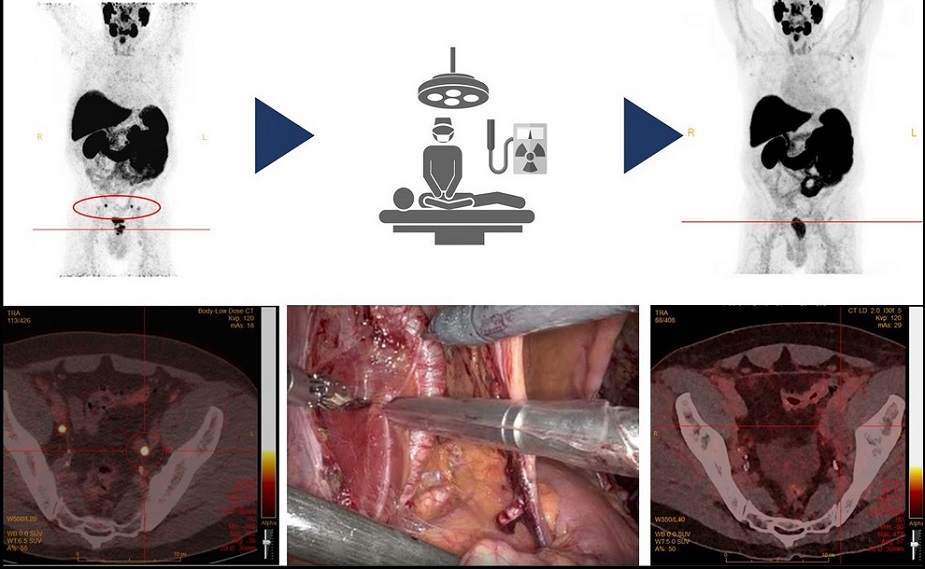
Now, a new study by researchers at the Radboud University Medical Centre (Nijmegen, Netherlands) has revealed that radioguided surgery is capable of detecting and extracting metastatic pelvic lymph nodes in patients newly diagnosed with prostate cancer. This technique focuses on the prostate-specific membrane antigen (PSMA), commonly overexpressed in prostate cancer cases, enhancing nodal staging and thereby aiding in forming treatment strategies for this crucial patient group. The study incorporated 20 newly diagnosed patients with prostate cancer, each with at least one lymph node suspected of metastasis based on a preoperative 18F-PSMA PET/CT scan. They underwent 111In-PSMA-radioguided surgery to remove the metastatic nodes, followed by a postoperative 18F-PSMA PET/CT scan confirming the successful excision of these lesions. This study was crucial in evaluating the safety and practicality of 111In-PSMA-radioguided surgery and its precision in identifying metastatic lymph nodes.

The study reported no adverse events linked to the 111In-PSMA-radioguided surgery. In the procedure, 29 out of 49 lesions were identified and excised, with 28 of them (97%) confirmed to be lymph node metastases. Additionally, 14 out of the 49 (29%) removed lymph nodes, which were not identified by the radioguided surgery, included two that had metastases. Although previous research has examined the feasibility of PSMA-radioguided pelvic lymph node surgery, this study is among the first to explore this method in a larger cohort of newly diagnosed patients. The results underscore the safety and feasibility of this innovative surgical technique. Importantly, each patient underwent postoperative imaging, a vital step in confirming the reliability of the outcomes.
“The current results demonstrate the great potential for radioguided surgery in prostate cancer and highlight the expanding role of molecular imaging at the operating room,” said Mark Rijpkema, PhD, principal investigator at the Radboud University Medical Centre. “Optimization of tracers and larger clinical trials may further improve surgical outcomes in the future by implementing both measurements of removed tissue, as well as real-time measurements during surgery.”
Related Links:
Radboud University Medical Centre


