New Radiotheranostic System Detects and Treats Ovarian Cancer Noninvasively
Posted on 04 Apr 2024
Ovarian cancer is the most lethal gynecological cancer, with less than a 30% five-year survival rate for those diagnosed in late stages. Despite surgery and platinum-based chemotherapy being the standard treatments, they have not significantly improved survival rates due to tumor recurrence and resistance to chemotherapy. Current serum-based biomarkers fail to correctly identify early-stage ovarian cancer, highlighting an urgent need for better detection methods and new targeted treatments to enhance patient survival rates. Now, a new radiotheranostic system offers non-invasive detection and treatment of ovarian cancer. This theranostic platform combines the highly specific huAR9.6 antibody with PET imaging and therapeutic radionuclides to provide a more personalized treatment that could potentially improve health outcomes for ovarian cancer patients.
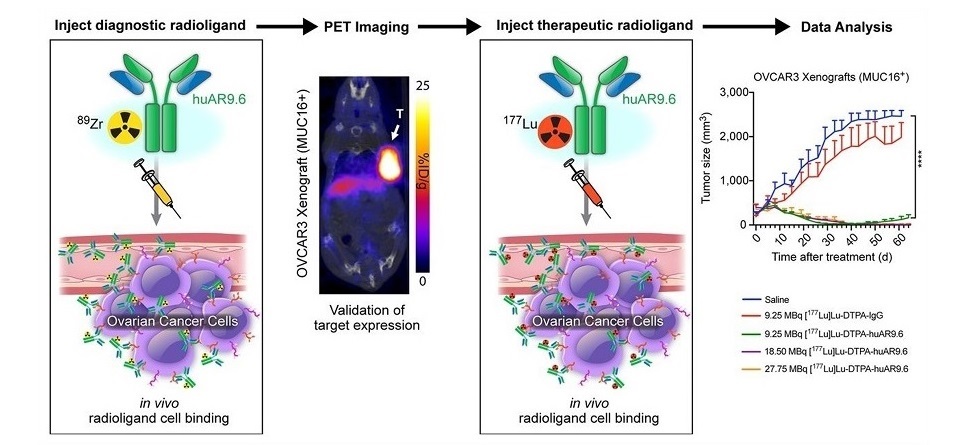
Previous studies have shown that the MUC16 protein is overexpressed in ovarian cancer patients, with elevated levels correlating with disease stage and tumor volume. The antibody huAR9.6 binds to a unique epitope that is influenced by truncated carbohydrate residues on MUC16. The MUC16 protein could be a potential target for tumor detection via immuno-PET imaging and treatment with radioimmunotherapy, according to findings of a study by researchers at the Memorial Sloan Kettering Cancer (MSK, New York, NY, USA). The study examined the diagnostic radiotracer 89Zr-DFO-huAR9.6 using vitro and in vivo via binding experiments, immuno-PET imaging, and biodistribution analysis on ovarian cancer mouse models.

Moreover, the therapeutic safety and efficacy of the radionuclide 177Lu-CHX-A″-DTPA-huAR9.6 were evaluated using ovarian xenografts. Immuno-PET imaging with 89Zr-DFO-huAR9.6 successfully identified MUC16 proteins, and in vivo tests confirmed the ability of 89Zr-DFO-huAR9.6 to distinguish varying MUC16 expression levels in ovarian cancer mouse models. Radioimmunotherapy studies with 177Lu-CHX-A″-DTPA-huAR9.6 showed promising results, including improved survival and strong antitumor response in models with high MUC16 expression. Hematologic toxicity was also determined to be transient in mice treated with 177Lu-CHX-A″-DTPA-huAR9.6.
“Immuno-PET imaging of MUC16 with this radiotheranostic pair may allow for noninvasive diagnosis and treatment monitoring of ovarian cancer lesions in patients,” said Kyeara Mack, PhD, postdoctoral fellow in the Lewis Lab at MSK. “This theranostic platform may be used to stratify and select patients who would benefit from the targeted radioimmunotherapy. In addition, it could also play a significant role in early ovarian cancer detection.”
Related Links:
MSK


