Imaging Software Improves Lung Diagnosis in Patients Allergic To Medical Contrast Dye
Posted on 21 May 2024
For up to 30% of patients who cannot use medical contrast dye due to allergies or other health conditions, diagnosing critical lung issues like pulmonary embolism can be delayed. This is because non-contrast dye imaging methods are less accurate and typically take longer to administer. Now, new imaging software has been developed to address this widespread challenge, offering such patients a more reliable and quicker diagnostic alternative.
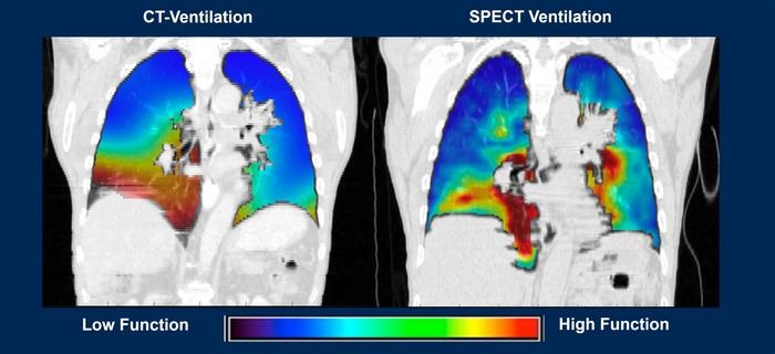
Developed at Corewell Health (Southfield, MI, USA), the software known as CT-Derived Functional Imaging, or CTFI, utilizes advanced computed tomography technology. It employs a complex mathematical approach integrated into a formula called the Integrated Jacobian Formulation, which swiftly calculates significant changes in lung volume as a patient breathes in and out. Furthermore, this method tracks changes in blood mass during the inhalation and exhalation phases, providing insights while the blood circulates through the lungs. This capability allows physicians and researchers to obtain consistent patient data, improving the accuracy of diagnoses and the precision of targeted treatments—all without the need for contrast dye. While this software is particularly beneficial for patients who cannot receive contrast dye, it also aids in the management of lung conditions such as chronic obstructive pulmonary disease (COPD) and cancer.

This innovative software has shown potential in minimizing radiation exposure to healthy lung areas adjacent to tumors during treatments. Additionally, it has proven effective in detecting pulmonary embolism by identifying changes in blood mass through a simple non-contrast CT scan during inhalation and exhalation. Recent findings demonstrate the software’s ability to predict disease progression in COPD patients over a decade, a feat that surpasses the capabilities of existing technologies. Researchers are now exploring how integrating a CTFI-based machine learning model with CT scans can enhance diagnostic accuracy, helping doctors better identify patients at risk of disease progression and ultimately improving survival rates for those with COPD.
“Ultimately, the goal is always to ensure doctors have the best tools available to them when treating patients,” said biomedical engineer Edward Castillo, Ph.D. “AI has the potential to further our work significantly in order to improve patient health and save lives.”
Related Links:
Corewell Health


