Game-Changing Technology Uses Live X-Ray Images for Guiding Endovascular Surgery
|
By MedImaging International staff writers Posted on 07 Jul 2022 |

Endovascular aneurysm repair (EVAR) is an alternative to open aortic surgery due to perceived advantages in patient survival, reduced post-operative complications and shorter hospital lengths of stay. Despite these potential advantages, there is still significant variability in pre-operative planning and sizing, problems associated with imprecise visualization and device positioning intra-operatively, and inconsistent patient outcomes. Now, a game-changing technology for vascular navigation aids in planning and guiding endovascular surgery and is simple to integrate with the existing imaging hardware that is already present in the hospital.
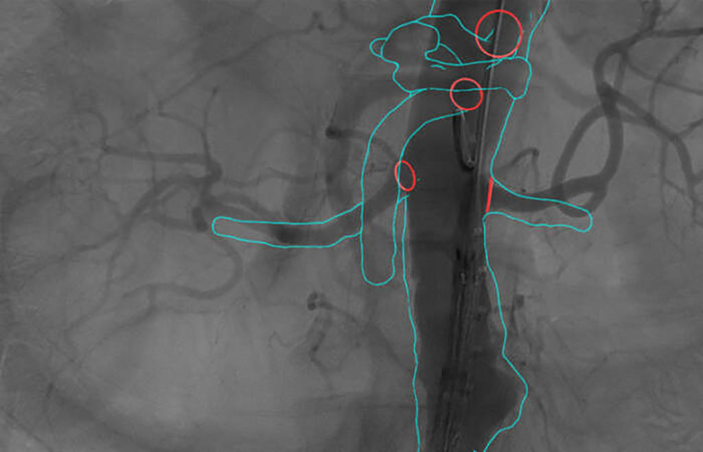
Cydar Medical’s (Cambridge, UK) Cydar EV is the first product from Cydar’s Intelligent Maps system. The patented computer vision automatically overlays the Map on the live X-ray imaging with exceptional accuracy and robustness. When guidewires and instruments deform the blood vessels, real-time imaging is used to update the Map to match the new, deformed anatomy. The result is an accurate, responsive 3D Map on the screen throughout a procedure.
During endovascular surgery, stiff guidewires often straighten, shorten and displace blood vessels. The surgeon uses grab handles positioned along virtual guide wires to adjust the shape of the 3D Map to match the real-time anatomy (non-rigid transformation). And, once adjusted, the system remembers that adjustment in 3D even when the X-ray set moves position. Toggling between the pre-operative map and the adjusted map helps the clinical team visualize how the anatomy has changed and position devices precisely. This reduces procedure length by 30-60 minutes in endovascular interventions and radiation exposure for clinical staff and patients is radically reduced, by 50% even in standard EVAR.
Cydar, in partnership with King’s College London (London, UK), has now initiated the ARIA Study: a randomized controlled trial to assess the clinical, technical and cost-effectiveness of a cloud-based, ARtificially Intelligent image fusion system in comparison to standard treatment to guide endovascular aortic aneurysm repair (ARIA). The randomized trial will enroll 340 patients in 10 sites across the UK with a clinical diagnosis of abdominal aortic or thoracoabdominal aortic aneurysm (AAA and TAAA respectively) suitable for endovascular treatment. The trial will follow patients for one year and assess the effect of Cydar EV Maps on clinical-, technical- and cost-effectiveness in comparison to standard treatment in endovascular aortic aneurysm repair, used for both standard and complex devices.
“Our central hypothesis is that digital technology - specifically cloud-computing and artificial intelligence (AI), can be used to assess and learn from large volumes of data to inform clinical decision making and has the potential to improve the predictability of individual patient outcomes and the consistency of outcomes in the NHS,” said Dr Rachel Clough, Principal Investigator of the ARIA Study and Clinical Senior Lecturer from King’s College London.
“Cydar EV Maps is a game-changing technology for vascular navigation. The ARIA study provides a unique opportunity to demonstrate the benefits like reduced procedure time and reduction to radiation exposure, although some of the more subtle benefits related to procedural quality and reduced operator fatigue may never be directly measured but are obvious as an operator,” said Dr. Simon Neequaye, Principal Investigator at the Liverpool University Hospital NHS Foundation Trust.
Related Links:
Cydar Medical
King’s College London
Latest Radiography News
- Routine Mammograms Could Predict Future Cardiovascular Disease in Women
- AI Detects Early Signs of Aging from Chest X-Rays
- X-Ray Breakthrough Captures Three Image-Contrast Types in Single Shot
- AI Generates Future Knee X-Rays to Predict Osteoarthritis Progression Risk
- AI Algorithm Uses Mammograms to Accurately Predict Cardiovascular Risk in Women
- AI Hybrid Strategy Improves Mammogram Interpretation
- AI Technology Predicts Personalized Five-Year Risk of Developing Breast Cancer
- RSNA AI Challenge Models Can Independently Interpret Mammograms
- New Technique Combines X-Ray Imaging and Radar for Safer Cancer Diagnosis
- New AI Tool Helps Doctors Read Chest X‑Rays Better
- Wearable X-Ray Imaging Detecting Fabric to Provide On-The-Go Diagnostic Scanning
- AI Helps Radiologists Spot More Lesions in Mammograms
- AI Detects Fatty Liver Disease from Chest X-Rays
- AI Detects Hidden Heart Disease in Existing CT Chest Scans
- Ultra-Lightweight AI Model Runs Without GPU to Break Barriers in Lung Cancer Diagnosis
- AI Radiology Tool Identifies Life-Threatening Conditions in Milliseconds

Channels
Radiography
view channel
Routine Mammograms Could Predict Future Cardiovascular Disease in Women
Mammograms are widely used to screen for breast cancer, but they may also contain overlooked clues about cardiovascular health. Calcium deposits in the arteries of the breast signal stiffening blood vessels,... Read more
AI Detects Early Signs of Aging from Chest X-Rays
Chronological age does not always reflect how fast the body is truly aging, and current biological age tests often rely on DNA-based markers that may miss early organ-level decline. Detecting subtle, age-related... Read moreMRI
view channel
New Material Boosts MRI Image Quality
Magnetic resonance imaging (MRI) is a cornerstone of modern diagnostics, yet certain deep or anatomically complex tissues, including delicate structures of the eye and orbit, remain difficult to visualize clearly.... Read more
AI Model Reads and Diagnoses Brain MRI in Seconds
Brain MRI scans are critical for diagnosing strokes, hemorrhages, and other neurological disorders, but interpreting them can take hours or even days due to growing demand and limited specialist availability.... Read moreMRI Scan Breakthrough to Help Avoid Risky Invasive Tests for Heart Patients
Heart failure patients often require right heart catheterization to assess how severely their heart is struggling to pump blood, a procedure that involves inserting a tube into the heart to measure blood... Read more
MRI Scans Reveal Signature Patterns of Brain Activity to Predict Recovery from TBI
Recovery after traumatic brain injury (TBI) varies widely, with some patients regaining full function while others are left with lasting disabilities. Prognosis is especially difficult to assess in patients... Read moreUltrasound
view channel
AI Model Accurately Detects Placenta Accreta in Pregnancy Before Delivery
Placenta accreta spectrum (PAS) is a life-threatening pregnancy complication in which the placenta abnormally attaches to the uterine wall. The condition is a leading cause of maternal mortality and morbidity... Read more
Portable Ultrasound Sensor to Enable Earlier Breast Cancer Detection
Breast cancer screening relies heavily on annual mammograms, but aggressive tumors can develop between scans, accounting for up to 30 percent of cases. These interval cancers are often diagnosed later,... Read more
Portable Imaging Scanner to Diagnose Lymphatic Disease in Real Time
Lymphatic disorders affect hundreds of millions of people worldwide and are linked to conditions ranging from limb swelling and organ dysfunction to birth defects and cancer-related complications.... Read more
Imaging Technique Generates Simultaneous 3D Color Images of Soft-Tissue Structure and Vasculature
Medical imaging tools often force clinicians to choose between speed, structural detail, and functional insight. Ultrasound is fast and affordable but typically limited to two-dimensional anatomy, while... Read moreNuclear Medicine
view channel
Radiopharmaceutical Molecule Marker to Improve Choice of Bladder Cancer Therapies
Targeted cancer therapies only work when tumor cells express the specific molecular structures they are designed to attack. In urothelial carcinoma, a common form of bladder cancer, the cell surface protein... Read more
Cancer “Flashlight” Shows Who Can Benefit from Targeted Treatments
Targeted cancer therapies can be highly effective, but only when a patient’s tumor expresses the specific protein the treatment is designed to attack. Determining this usually requires biopsies or advanced... Read moreGeneral/Advanced Imaging
view channel
AI Tool Offers Prognosis for Patients with Head and Neck Cancer
Oropharyngeal cancer is a form of head and neck cancer that can spread through lymph nodes, significantly affecting survival and treatment decisions. Current therapies often involve combinations of surgery,... Read more
New 3D Imaging System Addresses MRI, CT and Ultrasound Limitations
Medical imaging is central to diagnosing and managing injuries, cancer, infections, and chronic diseases, yet existing tools each come with trade-offs. Ultrasound, X-ray, CT, and MRI can be costly, time-consuming,... Read moreImaging IT
view channel
New Google Cloud Medical Imaging Suite Makes Imaging Healthcare Data More Accessible
Medical imaging is a critical tool used to diagnose patients, and there are billions of medical images scanned globally each year. Imaging data accounts for about 90% of all healthcare data1 and, until... Read more
Global AI in Medical Diagnostics Market to Be Driven by Demand for Image Recognition in Radiology
The global artificial intelligence (AI) in medical diagnostics market is expanding with early disease detection being one of its key applications and image recognition becoming a compelling consumer proposition... Read moreIndustry News
view channel
Nuclear Medicine Set for Continued Growth Driven by Demand for Precision Diagnostics
Clinical imaging services face rising demand for precise molecular diagnostics and targeted radiopharmaceutical therapy as cancer and chronic disease rates climb. A new market analysis projects rapid expansion... Read more


 Guided Devices.jpg)


