Photon Counting Detectors Promise Fast Color X-Ray Images
Posted on 26 Feb 2025
For many years, healthcare professionals have depended on traditional 2D X-rays to diagnose common bone fractures, though small fractures or soft tissue damage, such as cancers, can often be missed. MRI scans, while more effective, are expensive, time-consuming, and not always suitable for routine screening. Now, a new 3D technology promises to transform medical imaging, providing a faster, more accurate, and more affordable alternative to conventional diagnostic methods.
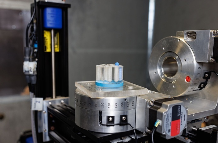
Researchers from the University of Houston (Houston, TX, USA) have demonstrated how photon-counting detectors combined with innovative algorithms enable more precise 3D visualization of various tissues and contrast agents. These detectors capture X-rays at multiple energy levels simultaneously, helping to differentiate materials within the body. Currently, X-rays used in medical facilities and industries capture incoming photons as a whole, similar to how white light contains all the colors, without separating them. While traditional X-rays can distinguish differences in density, such as bone from soft tissue, they can't identify the specific materials inside the body.

The photon-counting detectors developed by the team can separate X-ray photons based on their energy levels, much like a prism splits white light into its component colors. This ability enables the identification of specific materials, such as aluminum, plastic, iodine, or contrast agents like gadolinium used in medical imaging. This advancement could significantly improve cancer detection, for instance, by visualizing the accumulation of different contrast agents targeting a tumor and inflammation. Currently, while bright areas in images can be seen, it is often difficult to identify what they represent. This new technology, discussed in a paper featured on the cover of Journal of Medical Imaging, offers a clearer, more quantitative analysis that would allow clinicians to not only see what's inside the body but also identify the materials present and their quantities.
However, even with this enhanced detection, some materials share similar X-ray properties, making it difficult to distinguish more than two or three materials at once. This challenge is further complicated by errors in the detectors when separating photons by energy levels. To address this, the research team has developed a method to compensate for these detector distortions by calibrating the detectors using known materials. Once calibrated, the data can be processed using the novel algorithm to accurately decompose an image into its constituent materials. This multi-step process improves accuracy, using the same CT data collected. Although significant work remains before these advanced detectors can be used widely, the research team is collaborating with industry partners in Europe to develop larger versions of these detectors and refine their performance.
“We’re still in the research and development phase,” said Mini Das, Moores professor at UH’s College of Natural Sciences and Mathematics and Cullen College of Engineering, who developed the 3D solution. “Right now, the detectors are small, and we need to refine their measurement accuracy. But once we solve those challenges, we can begin testing in real-world medical and industrial settings.”


