Overhead Digital X-Ray Reduces Workflow Burdens
|
By MedImaging International staff writers Posted on 25 Oct 2021 |

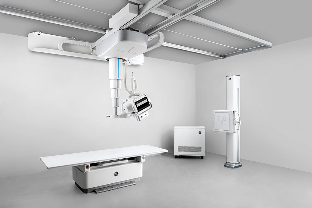
Image: The Definium Tempo radiology solution improve workflow ergonomics (Photo courtesy of GE Healthcare)
A new fixed overhead tube suspension (OTS) digital X-ray system leverages automation to help radiology departments deliver the best patient care possible.
The GE Healthcare (GE; Chicago, IL, USA) Definium Tempo is designed to act as an in-room command center, with a tube-mounted console providing all the functionality needed for patient selection, protocol selection, technique modification, and positioning setup, all without having to leave the patient’s side. Automated workflows and features such as auto positioning, auto centering, and auto tracking, automate the positioning of system components to maximize overall ergonomic operation for the technologist, while also helping to improve overall patient experience.
A collection of workflow enhancement tools formed by seamlessly combining a 3D video camera, computer vision, and video analytics help address positioning errors, poor image quality due to incorrect patient habitus selection, and ambiguities during image reading due to unique conditions. The GE Intelligent Workflow Suite includes Position Assist, Technique Assist, and Patient Snapshot to avoiding image retakes; produce more consistent images; increase technologists’ productivity; and improve clinical confidence by avoiding radiologist follow-up during diagnosis.
“I’ve been a technologist for over 17 years and using this system has been unlike anything I’ve experienced before; it’s a person inside the room with you at all times, it’s like you’re never working alone, it’s like a second technologist…” said X-ray technologist Nadia Dorson, of North Central Bronx Hospital (NY, USA). “The camera provides real-time information on the patient so you can make necessary adjustments. And everything is as light as a feather, even down to the grid, so I’m able to function better and am not encountering some of the day-to-day wear and tear I’ve experienced in the past.”
The daily challenges of heavy lifting, repetitive motions, uneasy patients, and long hours lead to more than 70% of technologists experiencing work-related injuries. Variability in patient positioning and exam set up can lead to extra dose with ‘repeat and reject’ rates (the need to retake images), as high as 25%. These pressures add additional burden to already strained radiology departments.
Related Links:
GE Healthcare
Latest Radiography News
- Routine Mammograms Could Predict Future Cardiovascular Disease in Women
- AI Detects Early Signs of Aging from Chest X-Rays
- X-Ray Breakthrough Captures Three Image-Contrast Types in Single Shot
- AI Generates Future Knee X-Rays to Predict Osteoarthritis Progression Risk
- AI Algorithm Uses Mammograms to Accurately Predict Cardiovascular Risk in Women
- AI Hybrid Strategy Improves Mammogram Interpretation
- AI Technology Predicts Personalized Five-Year Risk of Developing Breast Cancer
- RSNA AI Challenge Models Can Independently Interpret Mammograms
- New Technique Combines X-Ray Imaging and Radar for Safer Cancer Diagnosis
- New AI Tool Helps Doctors Read Chest X‑Rays Better
- Wearable X-Ray Imaging Detecting Fabric to Provide On-The-Go Diagnostic Scanning
- AI Helps Radiologists Spot More Lesions in Mammograms
- AI Detects Fatty Liver Disease from Chest X-Rays
- AI Detects Hidden Heart Disease in Existing CT Chest Scans
- Ultra-Lightweight AI Model Runs Without GPU to Break Barriers in Lung Cancer Diagnosis
- AI Radiology Tool Identifies Life-Threatening Conditions in Milliseconds

Channels
MRI
view channel
MRI Scans Reveal Signature Patterns of Brain Activity to Predict Recovery from TBI
Recovery after traumatic brain injury (TBI) varies widely, with some patients regaining full function while others are left with lasting disabilities. Prognosis is especially difficult to assess in patients... Read more
Novel Imaging Approach to Improve Treatment for Spinal Cord Injuries
Vascular dysfunction in the spinal cord contributes to multiple neurological conditions, including traumatic injuries and degenerative cervical myelopathy, where reduced blood flow can lead to progressive... Read more
AI-Assisted Model Enhances MRI Heart Scans
A cardiac MRI can reveal critical information about the heart’s function and any abnormalities, but traditional scans take 30 to 90 minutes and often suffer from poor image quality due to patient movement.... Read more
AI Model Outperforms Doctors at Identifying Patients Most At-Risk of Cardiac Arrest
Hypertrophic cardiomyopathy is one of the most common inherited heart conditions and a leading cause of sudden cardiac death in young individuals and athletes. While many patients live normal lives, some... Read moreUltrasound
view channel
Wearable Ultrasound Imaging System to Enable Real-Time Disease Monitoring
Chronic conditions such as hypertension and heart failure require close monitoring, yet today’s ultrasound imaging is largely confined to hospitals and short, episodic scans. This reactive model limits... Read more
Ultrasound Technique Visualizes Deep Blood Vessels in 3D Without Contrast Agents
Producing clear 3D images of deep blood vessels has long been difficult without relying on contrast agents, CT scans, or MRI. Standard ultrasound typically provides only 2D cross-sections, limiting clinicians’... Read moreNuclear Medicine
view channel
Cancer “Flashlight” Shows Who Can Benefit from Targeted Treatments
Targeted cancer therapies can be highly effective, but only when a patient’s tumor expresses the specific protein the treatment is designed to attack. Determining this usually requires biopsies or advanced... Read more
PET Imaging of Inflammation Predicts Recovery and Guides Therapy After Heart Attack
Acute myocardial infarction can trigger lasting heart damage, yet clinicians still lack reliable tools to identify which patients will regain function and which may develop heart failure.... Read more
Radiotheranostic Approach Detects, Kills and Reprograms Aggressive Cancers
Aggressive cancers such as osteosarcoma and glioblastoma often resist standard therapies, thrive in hostile tumor environments, and recur despite surgery, radiation, or chemotherapy. These tumors also... Read more
New Imaging Solution Improves Survival for Patients with Recurring Prostate Cancer
Detecting recurrent prostate cancer remains one of the most difficult challenges in oncology, as standard imaging methods such as bone scans and CT scans often fail to accurately locate small or early-stage tumors.... Read moreGeneral/Advanced Imaging
view channel
AI-Based Tool Predicts Future Cardiovascular Events in Angina Patients
Stable coronary artery disease is a common cause of chest pain, yet accurately identifying patients at the highest risk of future heart attacks or death remains difficult. Standard coronary CT scans show... Read more
AI-Based Tool Accelerates Detection of Kidney Cancer
Diagnosing kidney cancer depends on computed tomography scans, often using contrast agents to reveal abnormalities in kidney structure. Tumors are not always searched for deliberately, as many scans are... Read moreImaging IT
view channel
New Google Cloud Medical Imaging Suite Makes Imaging Healthcare Data More Accessible
Medical imaging is a critical tool used to diagnose patients, and there are billions of medical images scanned globally each year. Imaging data accounts for about 90% of all healthcare data1 and, until... Read more
Global AI in Medical Diagnostics Market to Be Driven by Demand for Image Recognition in Radiology
The global artificial intelligence (AI) in medical diagnostics market is expanding with early disease detection being one of its key applications and image recognition becoming a compelling consumer proposition... Read moreIndustry News
view channel
GE HealthCare and NVIDIA Collaboration to Reimagine Diagnostic Imaging
GE HealthCare (Chicago, IL, USA) has entered into a collaboration with NVIDIA (Santa Clara, CA, USA), expanding the existing relationship between the two companies to focus on pioneering innovation in... Read more
Patient-Specific 3D-Printed Phantoms Transform CT Imaging
New research has highlighted how anatomically precise, patient-specific 3D-printed phantoms are proving to be scalable, cost-effective, and efficient tools in the development of new CT scan algorithms... Read more
Siemens and Sectra Collaborate on Enhancing Radiology Workflows
Siemens Healthineers (Forchheim, Germany) and Sectra (Linköping, Sweden) have entered into a collaboration aimed at enhancing radiologists' diagnostic capabilities and, in turn, improving patient care... Read more


 Guided Devices.jpg)


