AI Tool Uses Chest X-Rays to Identify COVID-19 Patients Likely to Develop Life-Threatening Complications with 80% Accuracy
|
By MedImaging International staff writers Posted on 13 May 2021 |

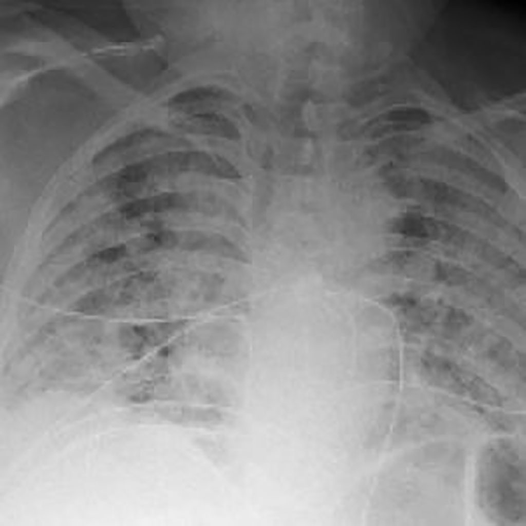
Illustration
Trained to see patterns by analyzing thousands of chest X-rays, a computer program predicted with up to 80% accuracy which patients with COVID-19 would develop life-threatening complications within four days.
Developed by researchers at NYU Grossman School of Medicine (New York, NY, USA), the program used several hundred gigabytes of data gleaned from 5,224 chest X-rays taken from 2,943 seriously ill patients infected with SARS-CoV-2, the virus behind the infections.
The authors of the study cited the “pressing need” for the ability to quickly predict which patients with COVID-19 are likely to have lethal complications so that treatment resources can best be matched to those at increased risk. For reasons not yet fully understood, the health of some patients with the disease suddenly worsens, requires intensive care, and increases their chances of dying. In a bid to address this need, the NYU Langone team fed not only X-ray information into their computer analysis, but also patients’ age, race, and gender, along with several vital signs and laboratory test results, including weight, body temperature, and blood immune cell levels. Also factored into their mathematical models, which can learn from examples, was the need for a mechanical ventilator and whether each patient survived (2,405) or died (538) from their infections.
Researchers then tested the predictive value of the software tool on 770 chest X-rays from 718 other patients admitted for COVID-19 through the emergency department at NYU Langone hospitals from March 3 to June 28, 2020. The computer program accurately predicted four out of five infected patients who required intensive care and mechanical ventilation and/or died within four days of admission.
A major advantage to machine intelligence programs such as this is that its accuracy can be tracked, updated, and improved with more data. The team plans to add more patient information as it becomes available and is also evaluating what additional clinical test results could be used to improve their test model. As part of further research, the team hopes to soon deploy NYU Langone’s COVID-19 classification test to emergency physicians and radiologists and is working with physicians to draft clinical guidelines for its use.
“Emergency room physicians and radiologists need effective tools like our program to quickly identify those patients with COVID-19 whose condition is most likely to deteriorate quickly so that healthcare providers can monitor them more closely and intervene earlier,” said study co-lead investigator Farah Shamout, PhD, an assistant professor in computer engineering at New York University’s campus in Abu Dhabi.
“We believe that our COVID-19 classification test represents the largest application of artificial intelligence in radiology to address some of the most urgent needs of patients and caregivers during the pandemic,” added Yiqiu “Artie” Shen, MS, a doctoral student at the NYU Center for Data Science.
Related Links:
NYU Grossman School of Medicine
Latest Radiography News
- Routine Mammograms Could Predict Future Cardiovascular Disease in Women
- AI Detects Early Signs of Aging from Chest X-Rays
- X-Ray Breakthrough Captures Three Image-Contrast Types in Single Shot
- AI Generates Future Knee X-Rays to Predict Osteoarthritis Progression Risk
- AI Algorithm Uses Mammograms to Accurately Predict Cardiovascular Risk in Women
- AI Hybrid Strategy Improves Mammogram Interpretation
- AI Technology Predicts Personalized Five-Year Risk of Developing Breast Cancer
- RSNA AI Challenge Models Can Independently Interpret Mammograms
- New Technique Combines X-Ray Imaging and Radar for Safer Cancer Diagnosis
- New AI Tool Helps Doctors Read Chest X‑Rays Better
- Wearable X-Ray Imaging Detecting Fabric to Provide On-The-Go Diagnostic Scanning
- AI Helps Radiologists Spot More Lesions in Mammograms
- AI Detects Fatty Liver Disease from Chest X-Rays
- AI Detects Hidden Heart Disease in Existing CT Chest Scans
- Ultra-Lightweight AI Model Runs Without GPU to Break Barriers in Lung Cancer Diagnosis
- AI Radiology Tool Identifies Life-Threatening Conditions in Milliseconds

Channels
Radiography
view channel
Routine Mammograms Could Predict Future Cardiovascular Disease in Women
Mammograms are widely used to screen for breast cancer, but they may also contain overlooked clues about cardiovascular health. Calcium deposits in the arteries of the breast signal stiffening blood vessels,... Read more
AI Detects Early Signs of Aging from Chest X-Rays
Chronological age does not always reflect how fast the body is truly aging, and current biological age tests often rely on DNA-based markers that may miss early organ-level decline. Detecting subtle, age-related... Read moreMRI
view channel
New Material Boosts MRI Image Quality
Magnetic resonance imaging (MRI) is a cornerstone of modern diagnostics, yet certain deep or anatomically complex tissues, including delicate structures of the eye and orbit, remain difficult to visualize clearly.... Read more
AI Model Reads and Diagnoses Brain MRI in Seconds
Brain MRI scans are critical for diagnosing strokes, hemorrhages, and other neurological disorders, but interpreting them can take hours or even days due to growing demand and limited specialist availability.... Read moreMRI Scan Breakthrough to Help Avoid Risky Invasive Tests for Heart Patients
Heart failure patients often require right heart catheterization to assess how severely their heart is struggling to pump blood, a procedure that involves inserting a tube into the heart to measure blood... Read more
MRI Scans Reveal Signature Patterns of Brain Activity to Predict Recovery from TBI
Recovery after traumatic brain injury (TBI) varies widely, with some patients regaining full function while others are left with lasting disabilities. Prognosis is especially difficult to assess in patients... Read moreUltrasound
view channel
Reusable Gel Pad Made from Tamarind Seed Could Transform Ultrasound Examinations
Ultrasound imaging depends on a conductive gel to eliminate air between the probe and the skin so sound waves can pass clearly into the body. While the imaging technology is fast, safe, and noninvasive,... Read more
AI Model Accurately Detects Placenta Accreta in Pregnancy Before Delivery
Placenta accreta spectrum (PAS) is a life-threatening pregnancy complication in which the placenta abnormally attaches to the uterine wall. The condition is a leading cause of maternal mortality and morbidity... Read moreNuclear Medicine
view channel
Radiopharmaceutical Molecule Marker to Improve Choice of Bladder Cancer Therapies
Targeted cancer therapies only work when tumor cells express the specific molecular structures they are designed to attack. In urothelial carcinoma, a common form of bladder cancer, the cell surface protein... Read more
Cancer “Flashlight” Shows Who Can Benefit from Targeted Treatments
Targeted cancer therapies can be highly effective, but only when a patient’s tumor expresses the specific protein the treatment is designed to attack. Determining this usually requires biopsies or advanced... Read moreGeneral/Advanced Imaging
view channel
AI Tool Offers Prognosis for Patients with Head and Neck Cancer
Oropharyngeal cancer is a form of head and neck cancer that can spread through lymph nodes, significantly affecting survival and treatment decisions. Current therapies often involve combinations of surgery,... Read more
New 3D Imaging System Addresses MRI, CT and Ultrasound Limitations
Medical imaging is central to diagnosing and managing injuries, cancer, infections, and chronic diseases, yet existing tools each come with trade-offs. Ultrasound, X-ray, CT, and MRI can be costly, time-consuming,... Read moreImaging IT
view channel
New Google Cloud Medical Imaging Suite Makes Imaging Healthcare Data More Accessible
Medical imaging is a critical tool used to diagnose patients, and there are billions of medical images scanned globally each year. Imaging data accounts for about 90% of all healthcare data1 and, until... Read more
Global AI in Medical Diagnostics Market to Be Driven by Demand for Image Recognition in Radiology
The global artificial intelligence (AI) in medical diagnostics market is expanding with early disease detection being one of its key applications and image recognition becoming a compelling consumer proposition... Read moreIndustry News
view channel
Nuclear Medicine Set for Continued Growth Driven by Demand for Precision Diagnostics
Clinical imaging services face rising demand for precise molecular diagnostics and targeted radiopharmaceutical therapy as cancer and chronic disease rates climb. A new market analysis projects rapid expansion... Read more


