Echocardiography Service Automatically Identifies Coronary Artery Disease
|
By MedImaging International staff writers Posted on 21 Jan 2021 |

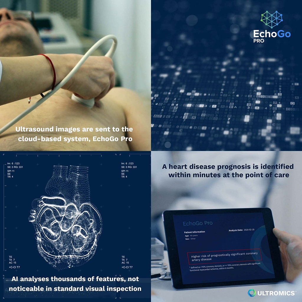
Image: The EchoGo Pro clinical workflows for CAD analysis (Photo courtesy of Ultromics)
An artificial intelligence (AI) powered, outcomes-driven, decision support tool improves the diagnostic accuracy of clinicians for coronary artery disease (CAD).
The Ultromics (Oxford, United Kingdom) EchoGo Pro is a fully automated solution for echocardiography and strain analysis. The cloud-based Software-as-a-Service (SaaS) platform uses AI to power the diagnostic pathway, providing near-instant reports without any need for physical software on site. EchoGo Pro complements EchoGo Core, a software application that measures standard cardiac parameters, including ejection fraction (EF), global longitudinal dtrain (GLS), and left ventricular (LV) volume, and also analyzes echocardiogram (ECG) cardiac parameters.
For Pro, the anonymized echocardiogram dataset is transferred and processed through the application’s workflow, and an auto-contouring algorithm places points around the LV that sufficiently capture the LV shape; these contours are then used for calculating geometric parameters. By automating ECG analysis as part of the cloud service, a complete accurate analysis can be delivered within minutes, and with zero variability, in a zero-click workflow, aiding identification of CAD. EchoGo Pro and EchoGo Core will be offered together in the EchoGo suite.
“Heart disease is the biggest killer globally, and this number is increasing daily. COVID-19 has placed an even greater pressure on cardiac care and looks likely to have lasting implications in terms of its impact on the heart,” said Ross Upton, MD, founder and CEO of Ultromics. “The healthcare industry needs to quickly pivot towards AI powered automation to reduce the time to diagnosis and improve patient care. We are making the EchoGo suite as complete and automated as possible to help clinicians rapidly assess disease and provide early, appropriate intervention.”
Cardiac ultrasound, or echocardiography, is a noninvasive diagnostic modality that can provide detailed hemodynamic information rapidly at the patient bedside. It was first adopted for diagnostic purposes in the 1960s as an inexpensive way to expedite diagnosis and management of imminently life-threatening cardiac disease, including pericardial tamponade, acute coronary syndrome (ACS), cardiomyopathy, pulmonary embolism (PE), and aortic dissection. Cardiac ultrasound can also differentiate shock states and guide resuscitative measures.
Related Links:
Ultromics
Latest Ultrasound News
- Wearable Ultrasound Imaging System to Enable Real-Time Disease Monitoring
- Ultrasound Technique Visualizes Deep Blood Vessels in 3D Without Contrast Agents
- Ultrasound Probe Images Entire Organ in 4D

- Disposable Ultrasound Patch Performs Better Than Existing Devices
- Non-Invasive Ultrasound-Based Tool Accurately Detects Infant Meningitis
- Breakthrough Deep Learning Model Enhances Handheld 3D Medical Imaging
- Pain-Free Breast Imaging System Performs One Minute Cancer Scan
- Wireless Chronic Pain Management Device to Reduce Need for Painkillers and Surgery
- New Medical Ultrasound Imaging Technique Enables ICU Bedside Monitoring
- New Incision-Free Technique Halts Growth of Debilitating Brain Lesions
- AI-Powered Lung Ultrasound Outperforms Human Experts in Tuberculosis Diagnosis
- AI Identifies Heart Valve Disease from Common Imaging Test
- Novel Imaging Method Enables Early Diagnosis and Treatment Monitoring of Type 2 Diabetes
- Ultrasound-Based Microscopy Technique to Help Diagnose Small Vessel Diseases
- Smart Ultrasound-Activated Immune Cells Destroy Cancer Cells for Extended Periods
- Tiny Magnetic Robot Takes 3D Scans from Deep Within Body
Channels
Radiography
view channel
Routine Mammograms Could Predict Future Cardiovascular Disease in Women
Mammograms are widely used to screen for breast cancer, but they may also contain overlooked clues about cardiovascular health. Calcium deposits in the arteries of the breast signal stiffening blood vessels,... Read more
AI Detects Early Signs of Aging from Chest X-Rays
Chronological age does not always reflect how fast the body is truly aging, and current biological age tests often rely on DNA-based markers that may miss early organ-level decline. Detecting subtle, age-related... Read moreMRI
view channel
MRI Scans Reveal Signature Patterns of Brain Activity to Predict Recovery from TBI
Recovery after traumatic brain injury (TBI) varies widely, with some patients regaining full function while others are left with lasting disabilities. Prognosis is especially difficult to assess in patients... Read more
Novel Imaging Approach to Improve Treatment for Spinal Cord Injuries
Vascular dysfunction in the spinal cord contributes to multiple neurological conditions, including traumatic injuries and degenerative cervical myelopathy, where reduced blood flow can lead to progressive... Read more
AI-Assisted Model Enhances MRI Heart Scans
A cardiac MRI can reveal critical information about the heart’s function and any abnormalities, but traditional scans take 30 to 90 minutes and often suffer from poor image quality due to patient movement.... Read more
AI Model Outperforms Doctors at Identifying Patients Most At-Risk of Cardiac Arrest
Hypertrophic cardiomyopathy is one of the most common inherited heart conditions and a leading cause of sudden cardiac death in young individuals and athletes. While many patients live normal lives, some... Read moreNuclear Medicine
view channel
PET Imaging of Inflammation Predicts Recovery and Guides Therapy After Heart Attack
Acute myocardial infarction can trigger lasting heart damage, yet clinicians still lack reliable tools to identify which patients will regain function and which may develop heart failure.... Read more
Radiotheranostic Approach Detects, Kills and Reprograms Aggressive Cancers
Aggressive cancers such as osteosarcoma and glioblastoma often resist standard therapies, thrive in hostile tumor environments, and recur despite surgery, radiation, or chemotherapy. These tumors also... Read more
New Imaging Solution Improves Survival for Patients with Recurring Prostate Cancer
Detecting recurrent prostate cancer remains one of the most difficult challenges in oncology, as standard imaging methods such as bone scans and CT scans often fail to accurately locate small or early-stage tumors.... Read moreGeneral/Advanced Imaging
view channel
AI-Based Tool Accelerates Detection of Kidney Cancer
Diagnosing kidney cancer depends on computed tomography scans, often using contrast agents to reveal abnormalities in kidney structure. Tumors are not always searched for deliberately, as many scans are... Read more
New Algorithm Dramatically Speeds Up Stroke Detection Scans
When patients arrive at emergency rooms with stroke symptoms, clinicians must rapidly determine whether the cause is a blood clot or a brain bleed, as treatment decisions depend on this distinction.... Read moreImaging IT
view channel
New Google Cloud Medical Imaging Suite Makes Imaging Healthcare Data More Accessible
Medical imaging is a critical tool used to diagnose patients, and there are billions of medical images scanned globally each year. Imaging data accounts for about 90% of all healthcare data1 and, until... Read more
Global AI in Medical Diagnostics Market to Be Driven by Demand for Image Recognition in Radiology
The global artificial intelligence (AI) in medical diagnostics market is expanding with early disease detection being one of its key applications and image recognition becoming a compelling consumer proposition... Read moreIndustry News
view channel
GE HealthCare and NVIDIA Collaboration to Reimagine Diagnostic Imaging
GE HealthCare (Chicago, IL, USA) has entered into a collaboration with NVIDIA (Santa Clara, CA, USA), expanding the existing relationship between the two companies to focus on pioneering innovation in... Read more
Patient-Specific 3D-Printed Phantoms Transform CT Imaging
New research has highlighted how anatomically precise, patient-specific 3D-printed phantoms are proving to be scalable, cost-effective, and efficient tools in the development of new CT scan algorithms... Read more
Siemens and Sectra Collaborate on Enhancing Radiology Workflows
Siemens Healthineers (Forchheim, Germany) and Sectra (Linköping, Sweden) have entered into a collaboration aimed at enhancing radiologists' diagnostic capabilities and, in turn, improving patient care... Read more


