Artificial Intelligence Add-On for `World’s First` Portable MR Imaging System Receives FDA Clearance
|
By MedImaging International staff writers Posted on 19 Jan 2021 |

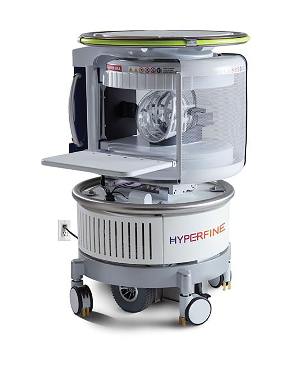
Image: Swoop Portable MR Imaging System (Photo courtesy of Hyperfine Research, Inc.)
Hyperfine Research, Inc. (Guilford, CT, USA) has received 510(k) clearance from the US FDA for its deep-learning image analysis software that measure brain structure and pathology in images acquired by the Swoop Portable MR Imaging System.
Magnetic Resonance Imaging (MRI) uses a magnetic field, radio waves, and a computer to produce detailed pictures of the body's internal structures that are clearer, more detailed, and more likely in some instances to identify and accurately characterize disease than other imaging methods. However, fixed MRI systems can be inconvenient and inaccessible for providers and patients, particularly when time is critical. Transport to the MR suite demands complicated scheduling coordination, moving patients, and, often, 4- to 6-hour patient backlogs — all of which compromise the utility of MRI as a diagnostic tool in time-sensitive settings, such as intensive care units and emergency rooms. Furthermore, high capital investments, electrical power needs, and significant maintenance requirements present barriers to adoption across all populations, acutely so for developing countries and rural geographies.
Hyperfine’s Swoop Portable MR Imaging System is designed to address the limitations of current imaging technologies and make MRI accessible anytime, anywhere, to any patient. Swoop wheels directly to the patient’s bedside, plugs into a standard electrical wall outlet, and is controlled by an Apple iPad. Designed as a complementary system to traditional MRIs at a fraction of the cost, images that display the internal structure of the head are captured by Swoop at the patient’s bedside, with results in minutes. Included as part of Swoop’s standard software package, Advanced AI Applications work with Swoop to transform the system into a true bedside clinical decision support platform for evaluating brain health and injury. In just minutes after Swoop scanning, Advanced AI Applications analyze and return annotated and segmented brain images, providing clinicians of all levels of expertise with quantitative markers for decision support and immediate feedback for diagnostic insight.
“With this powerful tool now built into the Swoop system, we are making MR imaging not only accessible at the bedside but making it easier for providers to move quickly from scan to a recommended course of treatment,” said Dr. Khan Siddiqui, Chief Medical Officer of Hyperfine. “The data provided by Hyperfine's deep learning software will liberate users of our MR technology by providing simple, accessible information in just minutes.”
Related Links:
Hyperfine Research, Inc.
Latest Industry News News
- GE HealthCare and NVIDIA Collaboration to Reimagine Diagnostic Imaging
- Patient-Specific 3D-Printed Phantoms Transform CT Imaging
- Siemens and Sectra Collaborate on Enhancing Radiology Workflows
- Bracco Diagnostics and ColoWatch Partner to Expand Availability CRC Screening Tests Using Virtual Colonoscopy
- Mindray Partners with TeleRay to Streamline Ultrasound Delivery
- Philips and Medtronic Partner on Stroke Care
- Siemens and Medtronic Enter into Global Partnership for Advancing Spine Care Imaging Technologies
- RSNA 2024 Technical Exhibits to Showcase Latest Advances in Radiology
- Bracco Collaborates with Arrayus on Microbubble-Assisted Focused Ultrasound Therapy for Pancreatic Cancer
- Innovative Collaboration to Enhance Ischemic Stroke Detection and Elevate Standards in Diagnostic Imaging
- RSNA 2024 Registration Opens
- Microsoft collaborates with Leading Academic Medical Systems to Advance AI in Medical Imaging
- GE HealthCare Acquires Intelligent Ultrasound Group’s Clinical Artificial Intelligence Business
- Bayer and Rad AI Collaborate on Expanding Use of Cutting Edge AI Radiology Operational Solutions
- Polish Med-Tech Company BrainScan to Expand Extensively into Foreign Markets
- Hologic Acquires UK-Based Breast Surgical Guidance Company Endomagnetics Ltd.
Channels
Radiography
view channel
Routine Mammograms Could Predict Future Cardiovascular Disease in Women
Mammograms are widely used to screen for breast cancer, but they may also contain overlooked clues about cardiovascular health. Calcium deposits in the arteries of the breast signal stiffening blood vessels,... Read more
AI Detects Early Signs of Aging from Chest X-Rays
Chronological age does not always reflect how fast the body is truly aging, and current biological age tests often rely on DNA-based markers that may miss early organ-level decline. Detecting subtle, age-related... Read moreMRI
view channel
MRI Scans Reveal Signature Patterns of Brain Activity to Predict Recovery from TBI
Recovery after traumatic brain injury (TBI) varies widely, with some patients regaining full function while others are left with lasting disabilities. Prognosis is especially difficult to assess in patients... Read more
Novel Imaging Approach to Improve Treatment for Spinal Cord Injuries
Vascular dysfunction in the spinal cord contributes to multiple neurological conditions, including traumatic injuries and degenerative cervical myelopathy, where reduced blood flow can lead to progressive... Read more
AI-Assisted Model Enhances MRI Heart Scans
A cardiac MRI can reveal critical information about the heart’s function and any abnormalities, but traditional scans take 30 to 90 minutes and often suffer from poor image quality due to patient movement.... Read more
AI Model Outperforms Doctors at Identifying Patients Most At-Risk of Cardiac Arrest
Hypertrophic cardiomyopathy is one of the most common inherited heart conditions and a leading cause of sudden cardiac death in young individuals and athletes. While many patients live normal lives, some... Read moreUltrasound
view channel
Wearable Ultrasound Imaging System to Enable Real-Time Disease Monitoring
Chronic conditions such as hypertension and heart failure require close monitoring, yet today’s ultrasound imaging is largely confined to hospitals and short, episodic scans. This reactive model limits... Read more
Ultrasound Technique Visualizes Deep Blood Vessels in 3D Without Contrast Agents
Producing clear 3D images of deep blood vessels has long been difficult without relying on contrast agents, CT scans, or MRI. Standard ultrasound typically provides only 2D cross-sections, limiting clinicians’... Read moreNuclear Medicine
view channel
Radiopharmaceutical Molecule Marker to Improve Choice of Bladder Cancer Therapies
Targeted cancer therapies only work when tumor cells express the specific molecular structures they are designed to attack. In urothelial carcinoma, a common form of bladder cancer, the cell surface protein... Read more
Cancer “Flashlight” Shows Who Can Benefit from Targeted Treatments
Targeted cancer therapies can be highly effective, but only when a patient’s tumor expresses the specific protein the treatment is designed to attack. Determining this usually requires biopsies or advanced... Read moreGeneral/Advanced Imaging
view channel
AI Tool Offers Prognosis for Patients with Head and Neck Cancer
Oropharyngeal cancer is a form of head and neck cancer that can spread through lymph nodes, significantly affecting survival and treatment decisions. Current therapies often involve combinations of surgery,... Read more
New 3D Imaging System Addresses MRI, CT and Ultrasound Limitations
Medical imaging is central to diagnosing and managing injuries, cancer, infections, and chronic diseases, yet existing tools each come with trade-offs. Ultrasound, X-ray, CT, and MRI can be costly, time-consuming,... Read moreImaging IT
view channel
New Google Cloud Medical Imaging Suite Makes Imaging Healthcare Data More Accessible
Medical imaging is a critical tool used to diagnose patients, and there are billions of medical images scanned globally each year. Imaging data accounts for about 90% of all healthcare data1 and, until... Read more


