Miniature Dosimeters Autonomously Monitor EMR Exposure
|
By MedImaging International staff writers Posted on 16 Jan 2020 |

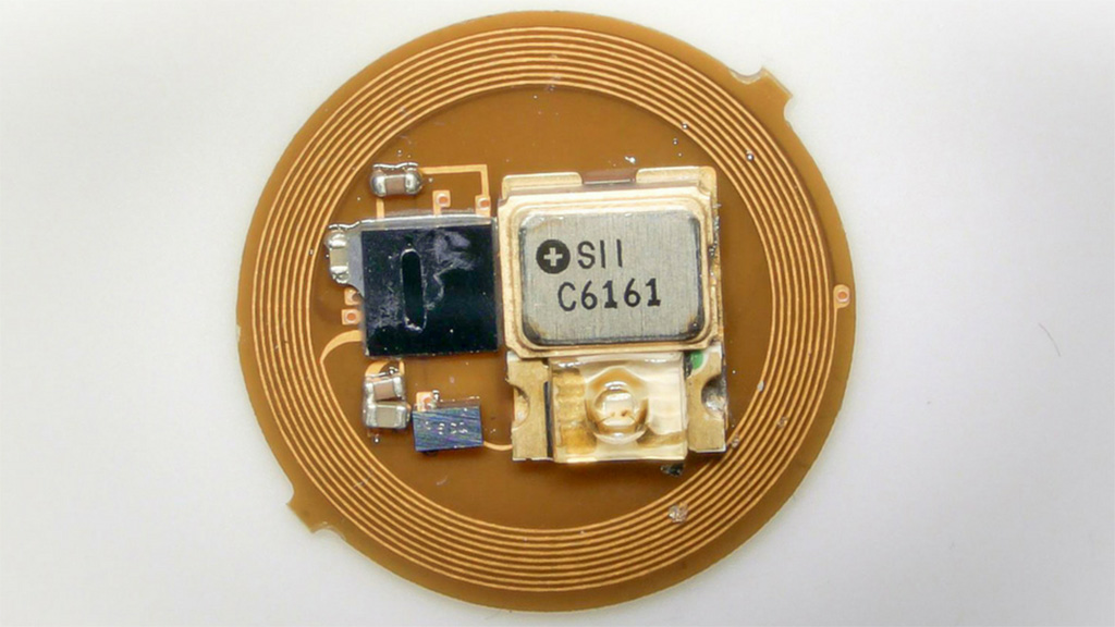
Image: A prototype autonomous EMR dosimeter (Photo courtesy of NU)
A millimeter-scale, ultra-low-power wireless digital platform provides continuous electromagnetic radiation (EMR) dosimetry for time-managed, wireless consumer devices.
Developed at Northwestern University (NU; Evanston, IL, USA) and the Korea Advanced Institute of Science and Technology (KAIST; Daejeon, Republic of Korea), the miniaturized digital dosimeter provides continuous EMR monitoring in an autonomous mode at one or multiple wavelengths simultaneously, transmitting the data over long-range wireless protocols to standard consumer devices. A single button cell battery powers the unit over a multiyear life span, enabled by the combined use of a light-powered, accumulation mode of detection and a light-adaptive, ultralow-power circuit design.
The dosimeter includes an accumulation detection module (ADM) for dosimetry and a Bluetooth low energy (BLE) system on a chip for wireless communication. A key feature is that the built-in ADM can directly measure continuous dose exposure without power consumption. As a result, it remains in an ultra-low sleep mode in the absence of light while continuously monitoring dosage via the ADM. When the dose exceeded a threshold, the device briefly wakes up to wirelessly transmit exposure data using BLE protocols to a smartphone, and resets the ADM and quickly return to sleep mode.
The ADM also includes a photodiode, supercapacitor, and a metal oxide semiconductor field-effect transistor (MOSFET). The miniaturized forms of the device have already been tested on sunglass clips, earrings, and wristbands for personalized EMR exposure detection. Field studies have shown that the dosimeter is extremely efficient in monitoring short-wavelength blue light from indoor lighting and display systems, as well as ultraviolet (UV), visible, and infrared (IR) radiation from the sun. The study was published on December 13, 2109, in Science Advances.
“The key feature of the ADM is that it directly measures exposure dose in a continuous fashion, without any power consumption. By contrast, conventional digital approaches approximate dose through computational time integration across a series of brief measurements of intensity, each performed using active, battery-powered electronics,” concluded lead author Kyeongha Kwon, PhD, of NU and KAIST, and colleagues. “Lack of interface ports and mechanical switches and the absence of need for battery replacement allow hermetic sealing of device for waterproof, sweat-resistant, and wear-resistant capabilities.”
Overexposure or underexposure to EMR can accumulate with latent consequences; where excessive exposure to UV and blue light from the sun or emissions of tanning beds and cellphones, can have associated health risks. For instance, repetitive keratinocyte damage from chronic exposure to UV is fundamental to cause skin cancer. The shorter wavelengths of the visible spectrum can generate reactive oxygen species (ROS) in the skin to cause DNA damage, hyperpigmentation and inflammation, alongside collagen and elastin degradation. Blue light can cause photochemical damage in retinal tissue to accelerate age-related maculopathy and modulate retinal control of the human circadian rhythm to suppress melatonin secretion.
Related Links:
Northwestern University
Korea Advanced Institute of Science and Technology
Latest Radiography News
- Routine Mammograms Could Predict Future Cardiovascular Disease in Women
- AI Detects Early Signs of Aging from Chest X-Rays
- X-Ray Breakthrough Captures Three Image-Contrast Types in Single Shot
- AI Generates Future Knee X-Rays to Predict Osteoarthritis Progression Risk
- AI Algorithm Uses Mammograms to Accurately Predict Cardiovascular Risk in Women
- AI Hybrid Strategy Improves Mammogram Interpretation
- AI Technology Predicts Personalized Five-Year Risk of Developing Breast Cancer
- RSNA AI Challenge Models Can Independently Interpret Mammograms
- New Technique Combines X-Ray Imaging and Radar for Safer Cancer Diagnosis
- New AI Tool Helps Doctors Read Chest X‑Rays Better
- Wearable X-Ray Imaging Detecting Fabric to Provide On-The-Go Diagnostic Scanning
- AI Helps Radiologists Spot More Lesions in Mammograms
- AI Detects Fatty Liver Disease from Chest X-Rays
- AI Detects Hidden Heart Disease in Existing CT Chest Scans
- Ultra-Lightweight AI Model Runs Without GPU to Break Barriers in Lung Cancer Diagnosis
- AI Radiology Tool Identifies Life-Threatening Conditions in Milliseconds

Channels
MRI
view channel
MRI Scans Reveal Signature Patterns of Brain Activity to Predict Recovery from TBI
Recovery after traumatic brain injury (TBI) varies widely, with some patients regaining full function while others are left with lasting disabilities. Prognosis is especially difficult to assess in patients... Read more
Novel Imaging Approach to Improve Treatment for Spinal Cord Injuries
Vascular dysfunction in the spinal cord contributes to multiple neurological conditions, including traumatic injuries and degenerative cervical myelopathy, where reduced blood flow can lead to progressive... Read more
AI-Assisted Model Enhances MRI Heart Scans
A cardiac MRI can reveal critical information about the heart’s function and any abnormalities, but traditional scans take 30 to 90 minutes and often suffer from poor image quality due to patient movement.... Read more
AI Model Outperforms Doctors at Identifying Patients Most At-Risk of Cardiac Arrest
Hypertrophic cardiomyopathy is one of the most common inherited heart conditions and a leading cause of sudden cardiac death in young individuals and athletes. While many patients live normal lives, some... Read moreUltrasound
view channel
Wearable Ultrasound Imaging System to Enable Real-Time Disease Monitoring
Chronic conditions such as hypertension and heart failure require close monitoring, yet today’s ultrasound imaging is largely confined to hospitals and short, episodic scans. This reactive model limits... Read more
Ultrasound Technique Visualizes Deep Blood Vessels in 3D Without Contrast Agents
Producing clear 3D images of deep blood vessels has long been difficult without relying on contrast agents, CT scans, or MRI. Standard ultrasound typically provides only 2D cross-sections, limiting clinicians’... Read moreNuclear Medicine
view channel
PET Imaging of Inflammation Predicts Recovery and Guides Therapy After Heart Attack
Acute myocardial infarction can trigger lasting heart damage, yet clinicians still lack reliable tools to identify which patients will regain function and which may develop heart failure.... Read more
Radiotheranostic Approach Detects, Kills and Reprograms Aggressive Cancers
Aggressive cancers such as osteosarcoma and glioblastoma often resist standard therapies, thrive in hostile tumor environments, and recur despite surgery, radiation, or chemotherapy. These tumors also... Read more
New Imaging Solution Improves Survival for Patients with Recurring Prostate Cancer
Detecting recurrent prostate cancer remains one of the most difficult challenges in oncology, as standard imaging methods such as bone scans and CT scans often fail to accurately locate small or early-stage tumors.... Read moreGeneral/Advanced Imaging
view channel
AI-Based Tool Accelerates Detection of Kidney Cancer
Diagnosing kidney cancer depends on computed tomography scans, often using contrast agents to reveal abnormalities in kidney structure. Tumors are not always searched for deliberately, as many scans are... Read more
New Algorithm Dramatically Speeds Up Stroke Detection Scans
When patients arrive at emergency rooms with stroke symptoms, clinicians must rapidly determine whether the cause is a blood clot or a brain bleed, as treatment decisions depend on this distinction.... Read moreImaging IT
view channel
New Google Cloud Medical Imaging Suite Makes Imaging Healthcare Data More Accessible
Medical imaging is a critical tool used to diagnose patients, and there are billions of medical images scanned globally each year. Imaging data accounts for about 90% of all healthcare data1 and, until... Read more
Global AI in Medical Diagnostics Market to Be Driven by Demand for Image Recognition in Radiology
The global artificial intelligence (AI) in medical diagnostics market is expanding with early disease detection being one of its key applications and image recognition becoming a compelling consumer proposition... Read moreIndustry News
view channel
GE HealthCare and NVIDIA Collaboration to Reimagine Diagnostic Imaging
GE HealthCare (Chicago, IL, USA) has entered into a collaboration with NVIDIA (Santa Clara, CA, USA), expanding the existing relationship between the two companies to focus on pioneering innovation in... Read more
Patient-Specific 3D-Printed Phantoms Transform CT Imaging
New research has highlighted how anatomically precise, patient-specific 3D-printed phantoms are proving to be scalable, cost-effective, and efficient tools in the development of new CT scan algorithms... Read more
Siemens and Sectra Collaborate on Enhancing Radiology Workflows
Siemens Healthineers (Forchheim, Germany) and Sectra (Linköping, Sweden) have entered into a collaboration aimed at enhancing radiologists' diagnostic capabilities and, in turn, improving patient care... Read more


