Handheld Device Aids Diagnosis of Skin Cancer
|
By MedImaging International staff writers Posted on 01 Jul 2019 |

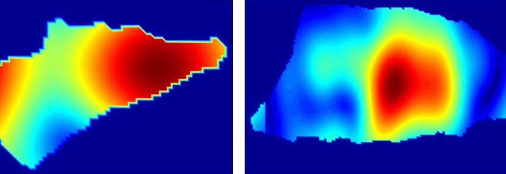
Image: Reflectivity patterns can distinguish between basal cell carcinoma (L) and squamous cell carcinoma (R) (Photo courtesy of Stevens Institute of Technology).
A new study shows how ultra-high-resolution millimeter-wave imaging (UH-MMWI) can be used to detects skin lesions and determine if they are cancerous or benign.
Developed by researchers at the Stevens Institute of Technology (Hoboken, NJ, USA) and Hackensack University Medical Center (NJ, USA), the new technology can realize three-dimensional (3D) high-contrast images of the skin by taking advantage of intrinsic dielectric contrasts between normal and malignant skin tissues. The system works in the 98 GHz bandwidth, which is also used in cellphones and airport security scanners. As cancerous tumors reflect around 40% more calibrated energy than healthy tissue, it is possible to identify diseased tissue by searching for reflectivity hotspots.
A programmable measurement platform automatically scan the tissues across a rectangular aperture plane, while a frequency domain imaging algorithm processes the recorded signals and generates an image of the cancerous tissue. To test the device, the researchers imaged 21 non-melanoma skin cancer specimens and compared them with histopathology. The results revealed a high correlation between MMWI and histological images, with reflectivity values statistically significant higher for cancerous areas compared to normal areas. The study was published on March 4, 2019, in IEEE Transactions on Medical Imaging.
“This technology can be used for rapidly detecting skin cancer. That's a major step forward toward our ultimate goal of developing a handheld device, which would be safe to use directly on the skin for an almost instant diagnostic reading of specific kinds of skin cancer, including lethal melanomas,” said senior author Negar Tavassolian, PhD, director of the Stevens Bio-Electromagnetics Laboratory. “We could place these devices in pharmacies, so people can get checked out and go to a doctor for a follow-up if necessary. People won't need to wait weeks to get results, and that will save lives.”
According to the researchers, it should be possible to keep manufacturing costs below USD 1,000, even at low production volumes, which is similar to the cost of current optical magnifying tools already used by dermatologists, and an order of magnitude cheaper than laser-based imaging tools, which also tend to be slower, bulkier, and less accurate than millimeter-wave scanners.
Related Links:
Stevens Institute of Technology
Hackensack University Medical Center
Latest General/Advanced Imaging News
- CT Colonography Beats Stool DNA Testing for Colon Cancer Screening
- First-Of-Its-Kind Wearable Device Offers Revolutionary Alternative to CT Scans
- AI-Based CT Scan Analysis Predicts Early-Stage Kidney Damage Due to Cancer Treatments
- CT-Based Deep Learning-Driven Tool to Enhance Liver Cancer Diagnosis
- AI-Powered Imaging System Improves Lung Cancer Diagnosis
- AI Model Significantly Enhances Low-Dose CT Capabilities
- Ultra-Low Dose CT Aids Pneumonia Diagnosis in Immunocompromised Patients
- AI Reduces CT Lung Cancer Screening Workload by Almost 80%
- Cutting-Edge Technology Combines Light and Sound for Real-Time Stroke Monitoring
- AI System Detects Subtle Changes in Series of Medical Images Over Time
- New CT Scan Technique to Improve Prognosis and Treatments for Head and Neck Cancers
- World’s First Mobile Whole-Body CT Scanner to Provide Diagnostics at POC
- Comprehensive CT Scans Could Identify Atherosclerosis Among Lung Cancer Patients
- AI Improves Detection of Colorectal Cancer on Routine Abdominopelvic CT Scans
- Super-Resolution Technology Enhances Clinical Bone Imaging to Predict Osteoporotic Fracture Risk
- AI-Powered Abdomen Map Enables Early Cancer Detection
Channels
Radiography
view channel
AI Radiology Tool Identifies Life-Threatening Conditions in Milliseconds
Radiology is emerging as one of healthcare’s most pressing bottlenecks. By 2033, the U.S. could face a shortage of up to 42,000 radiologists, even as imaging volumes grow by 5% annually.... Read more
Machine Learning Algorithm Identifies Cardiovascular Risk from Routine Bone Density Scans
A new study published in the Journal of Bone and Mineral Research reveals that an automated machine learning program can predict the risk of cardiovascular events and falls or fractures by analyzing bone... Read more
AI Improves Early Detection of Interval Breast Cancers
Interval breast cancers, which occur between routine screenings, are easier to treat when detected earlier. Early detection can reduce the need for aggressive treatments and improve the chances of better outcomes.... Read more
World's Largest Class Single Crystal Diamond Radiation Detector Opens New Possibilities for Diagnostic Imaging
Diamonds possess ideal physical properties for radiation detection, such as exceptional thermal and chemical stability along with a quick response time. Made of carbon with an atomic number of six, diamonds... Read moreMRI
view channel
New MRI Technique Reveals Hidden Heart Issues
Traditional exercise stress tests conducted within an MRI machine require patients to lie flat, a position that artificially improves heart function by increasing stroke volume due to gravity-driven blood... Read more
Shorter MRI Exam Effectively Detects Cancer in Dense Breasts
Women with extremely dense breasts face a higher risk of missed breast cancer diagnoses, as dense glandular and fibrous tissue can obscure tumors on mammograms. While breast MRI is recommended for supplemental... Read moreUltrasound
view channel
New Medical Ultrasound Imaging Technique Enables ICU Bedside Monitoring
Ultrasound computed tomography (USCT) presents a safer alternative to imaging techniques like X-ray computed tomography (commonly known as CT or “CAT” scans) because it does not produce ionizing radiation.... Read more
New Incision-Free Technique Halts Growth of Debilitating Brain Lesions
Cerebral cavernous malformations (CCMs), also known as cavernomas, are abnormal clusters of blood vessels that can grow in the brain, spinal cord, or other parts of the body. While most cases remain asymptomatic,... Read moreNuclear Medicine
view channel
New Imaging Approach Could Reduce Need for Biopsies to Monitor Prostate Cancer
Prostate cancer is the second leading cause of cancer-related death among men in the United States. However, the majority of older men diagnosed with prostate cancer have slow-growing, low-risk forms of... Read more
Novel Radiolabeled Antibody Improves Diagnosis and Treatment of Solid Tumors
Interleukin-13 receptor α-2 (IL13Rα2) is a cell surface receptor commonly found in solid tumors such as glioblastoma, melanoma, and breast cancer. It is minimally expressed in normal tissues, making it... Read moreImaging IT
view channel
New Google Cloud Medical Imaging Suite Makes Imaging Healthcare Data More Accessible
Medical imaging is a critical tool used to diagnose patients, and there are billions of medical images scanned globally each year. Imaging data accounts for about 90% of all healthcare data1 and, until... Read more
Global AI in Medical Diagnostics Market to Be Driven by Demand for Image Recognition in Radiology
The global artificial intelligence (AI) in medical diagnostics market is expanding with early disease detection being one of its key applications and image recognition becoming a compelling consumer proposition... Read moreIndustry News
view channel
GE HealthCare and NVIDIA Collaboration to Reimagine Diagnostic Imaging
GE HealthCare (Chicago, IL, USA) has entered into a collaboration with NVIDIA (Santa Clara, CA, USA), expanding the existing relationship between the two companies to focus on pioneering innovation in... Read more
Patient-Specific 3D-Printed Phantoms Transform CT Imaging
New research has highlighted how anatomically precise, patient-specific 3D-printed phantoms are proving to be scalable, cost-effective, and efficient tools in the development of new CT scan algorithms... Read more
Siemens and Sectra Collaborate on Enhancing Radiology Workflows
Siemens Healthineers (Forchheim, Germany) and Sectra (Linköping, Sweden) have entered into a collaboration aimed at enhancing radiologists' diagnostic capabilities and, in turn, improving patient care... Read more


.jpeg)


