Super-Resolution Technology Enhances Clinical Bone Imaging to Predict Osteoporotic Fracture Risk
Posted on 12 Feb 2025
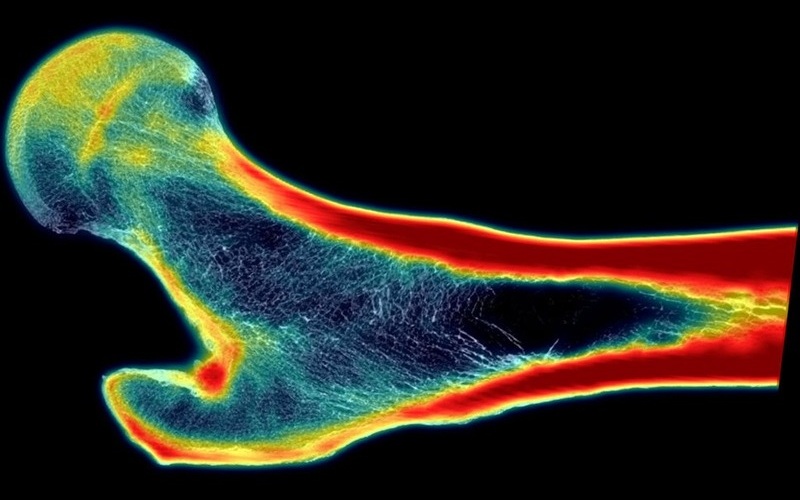
Osteoporotic fractures in the elderly represent a significant and widespread issue. Half of all women over the age of 55 will experience some form of fracture during their lifetime. Among the millions of individuals who suffer a hip fracture each year, 50% will never regain the ability to walk independently, and 20% will die within a year. Engineers and orthopedic clinicians are working to identify individuals at risk, as there are interventions available that can strengthen bones, but success depends on identifying who is most vulnerable. Currently, X-rays and CT scans are the most common methods used to assess fracture risk, but the resolution of both imaging techniques is too low to accurately assess the structural strength of the bone. Now, a new technology that improves clinical bone imaging and reduces osteoporotic fractures in the elderly uses artificial intelligence (AI) to generate super-resolution images that reveal the inner structures of bones in greater detail, helping to better assess fracture risk.
In their study, researchers at Southwest Research Institute (SwRI, San Antonio, TX, USA) discovered that in nearly all cases, super-resolution (SR)-enhanced images were more accurate than X-rays or CT scans at quantifying bone characteristics related to strength and fracture risk. The findings, published in the journal Bone, highlight the use of super-resolution technology, a type of AI that enhances the sharpness and detail of low-quality images. SwRI’s deep-learning SR method teaches the AI to recognize complex patterns and textures by exposing it to multiple examples, enabling it to predict what a higher-resolution image of the bone would look like based on low-resolution data.

The researchers have proposed integrating SR technology into medical imaging equipment, which would provide clinicians with both raw medical images, such as X-rays, and enhanced images processed with AI. These resulting images would be clearer and more detailed, potentially accompanied by additional diagnostics that could assist physicians in making more informed medical decisions. With access to high-resolution image data, various tools are available to predict the actual strength of the bone. Clinicians would be able to assess whether the bones are at risk of fracture and determine whether intervention is necessary to prevent injury. Once fracture risk is identified, doctors can recommend medications or exercises to strengthen vulnerable bones and reduce the likelihood of injury.
"Everybody falls. Everyone over the age of 55, and particularly those over 65, will fall at some point, so understanding bone strength for this population is important,” said SwRI’s Dr. Lance Frazer, the study’s lead author. “A lot can be done in terms of fall prevention. However, the imminent question remains: when you fall, are you at risk for a fracture? Knowing this is critical.”
Related Links:
SwRI


