New AI-Powered Lung Imaging Solution Launched at RSNA 2018
|
By MedImaging International staff writers Posted on 26 Nov 2018 |

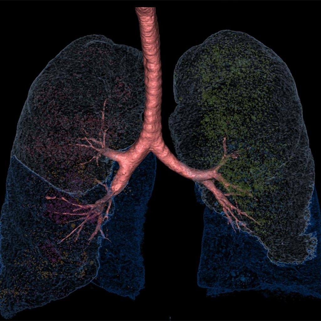
Image: LungPrint Discovery is designed as an artificial intelligence (AI)-powered lung analysis solution for radiologists (Photo courtesy of VIDA Diagnostics).
VIDA Diagnostics, Inc. (Coralville, IA, USA), a pulmonary imaging analytics company, launched LungPrint Discovery, an artificial intelligence (AI)-powered lung analysis solution for radiologists, at the Radiological Society of North America's (RSNA) 104th Scientific Assembly and Annual Meeting, held November 25-30 in Chicago, Ill., USA.
VIDA is focused on transforming pulmonary care through intelligence. Powered by a combination of AI and quality-controlled image analysis services, VIDA's solution aims to provide greater precision and personalization across a range of lung diseases, including cancer, emphysema, airway obstructive diseases, asthma and interstitial lung disease. Like a fingerprint, each lung is unique. The LungPrint family of products, starting with LungPrint Discovery, aims to reveal each patient's unique lung profile in a manner that is clinically meaningful and efficient, helping VIDA deliver on its mission to transform lung care through information and intelligence.
LungPrint Discovery, a part of the VIDA|vision Software Suite, provides fully automatic quantification of lung physiology and function, including both tissue and airway analysis. LungPrint Discovery also features an airway visualization called "Hyperion View" with the potential to significantly accelerate airway reading and interpreting the complex airway anatomy.
"I've had the opportunity to preview VIDA's LungPrint Discovery and it is a game-changer for the reporting of thoracic CT scans," said Dr. John Newell, Professor of Radiology at the University of Iowa. "It will empower radiologists to provide a richer set of quantitative CT lung information with a state-of-the-art CT report for the referring physician. The new airway display is remarkable."
"We are pleased to serve radiologists with a product that delivers on the promises of AI, namely greater and proven clinical precision in tandem with efficiency gains," said Susan A. Wood, PhD, CEO of VIDA. "Our solution continually evolves and is designed with leading clinicians and clinically validated through large-scale trials. With seamless integration into the radiology workflow, our best-in-class solution can be used with a broadening audience and make a greater impact on pulmonary patient care."
Related Links:
VIDA Diagnostics
Latest RSNA 2018 News
- Agfa Brings Intelligent Radiography to RSNA 2018
- Carestream Displays Several IT Offerings at Radiology Congress
- Double Black Imaging Announces Expanded Clinical LCD Line at Trade Show
- Lunit Unveils AI-Based Mammography Solution at RSNA 2018
- Guerbet Showcases Ongoing Collaboration with IBM Watson at Radiology Trade Fair
- Fujifilm Showcases New DR Detectors and AI Initiative in Chicago
- M*Modal Launches Cloud-Based Version of AI-Powered Reporting Solution
- MDW Unveils First Radiology Blockchain Platform at RSNA 2018
- Teledyne DALSA Displays Xineos Family of CMOS X-ray Detectors
- SuperSonic Imagine Showcases New Ultrasound System at RSNA 2018
- CIVCO Medical Solutions Introduces Next-Generation of Ultrasound Accessories
- Subtle Medical Showcases Al for PET and MRI Scans at RSNA 2018
- Philips Launches New Platform to Enable Development of AI Assets
Channels
Radiography
view channel
Routine Mammograms Could Predict Future Cardiovascular Disease in Women
Mammograms are widely used to screen for breast cancer, but they may also contain overlooked clues about cardiovascular health. Calcium deposits in the arteries of the breast signal stiffening blood vessels,... Read more
AI Detects Early Signs of Aging from Chest X-Rays
Chronological age does not always reflect how fast the body is truly aging, and current biological age tests often rely on DNA-based markers that may miss early organ-level decline. Detecting subtle, age-related... Read moreMRI
view channel
MRI Scans Reveal Signature Patterns of Brain Activity to Predict Recovery from TBI
Recovery after traumatic brain injury (TBI) varies widely, with some patients regaining full function while others are left with lasting disabilities. Prognosis is especially difficult to assess in patients... Read more
Novel Imaging Approach to Improve Treatment for Spinal Cord Injuries
Vascular dysfunction in the spinal cord contributes to multiple neurological conditions, including traumatic injuries and degenerative cervical myelopathy, where reduced blood flow can lead to progressive... Read more
AI-Assisted Model Enhances MRI Heart Scans
A cardiac MRI can reveal critical information about the heart’s function and any abnormalities, but traditional scans take 30 to 90 minutes and often suffer from poor image quality due to patient movement.... Read more
AI Model Outperforms Doctors at Identifying Patients Most At-Risk of Cardiac Arrest
Hypertrophic cardiomyopathy is one of the most common inherited heart conditions and a leading cause of sudden cardiac death in young individuals and athletes. While many patients live normal lives, some... Read moreUltrasound
view channel
Wearable Ultrasound Imaging System to Enable Real-Time Disease Monitoring
Chronic conditions such as hypertension and heart failure require close monitoring, yet today’s ultrasound imaging is largely confined to hospitals and short, episodic scans. This reactive model limits... Read more
Ultrasound Technique Visualizes Deep Blood Vessels in 3D Without Contrast Agents
Producing clear 3D images of deep blood vessels has long been difficult without relying on contrast agents, CT scans, or MRI. Standard ultrasound typically provides only 2D cross-sections, limiting clinicians’... Read moreNuclear Medicine
view channel
PET Imaging of Inflammation Predicts Recovery and Guides Therapy After Heart Attack
Acute myocardial infarction can trigger lasting heart damage, yet clinicians still lack reliable tools to identify which patients will regain function and which may develop heart failure.... Read more
Radiotheranostic Approach Detects, Kills and Reprograms Aggressive Cancers
Aggressive cancers such as osteosarcoma and glioblastoma often resist standard therapies, thrive in hostile tumor environments, and recur despite surgery, radiation, or chemotherapy. These tumors also... Read more
New Imaging Solution Improves Survival for Patients with Recurring Prostate Cancer
Detecting recurrent prostate cancer remains one of the most difficult challenges in oncology, as standard imaging methods such as bone scans and CT scans often fail to accurately locate small or early-stage tumors.... Read moreGeneral/Advanced Imaging
view channel
AI-Based Tool Predicts Future Cardiovascular Events in Angina Patients
Stable coronary artery disease is a common cause of chest pain, yet accurately identifying patients at the highest risk of future heart attacks or death remains difficult. Standard coronary CT scans show... Read more
AI-Based Tool Accelerates Detection of Kidney Cancer
Diagnosing kidney cancer depends on computed tomography scans, often using contrast agents to reveal abnormalities in kidney structure. Tumors are not always searched for deliberately, as many scans are... Read moreImaging IT
view channel
New Google Cloud Medical Imaging Suite Makes Imaging Healthcare Data More Accessible
Medical imaging is a critical tool used to diagnose patients, and there are billions of medical images scanned globally each year. Imaging data accounts for about 90% of all healthcare data1 and, until... Read more
Global AI in Medical Diagnostics Market to Be Driven by Demand for Image Recognition in Radiology
The global artificial intelligence (AI) in medical diagnostics market is expanding with early disease detection being one of its key applications and image recognition becoming a compelling consumer proposition... Read moreIndustry News
view channel
GE HealthCare and NVIDIA Collaboration to Reimagine Diagnostic Imaging
GE HealthCare (Chicago, IL, USA) has entered into a collaboration with NVIDIA (Santa Clara, CA, USA), expanding the existing relationship between the two companies to focus on pioneering innovation in... Read more
Patient-Specific 3D-Printed Phantoms Transform CT Imaging
New research has highlighted how anatomically precise, patient-specific 3D-printed phantoms are proving to be scalable, cost-effective, and efficient tools in the development of new CT scan algorithms... Read more
Siemens and Sectra Collaborate on Enhancing Radiology Workflows
Siemens Healthineers (Forchheim, Germany) and Sectra (Linköping, Sweden) have entered into a collaboration aimed at enhancing radiologists' diagnostic capabilities and, in turn, improving patient care... Read more


 Guided Devices.jpg)


