VR Tool Increases Efficiency of Brain Scan Analysis
|
By MedImaging International staff writers Posted on 06 Sep 2018 |

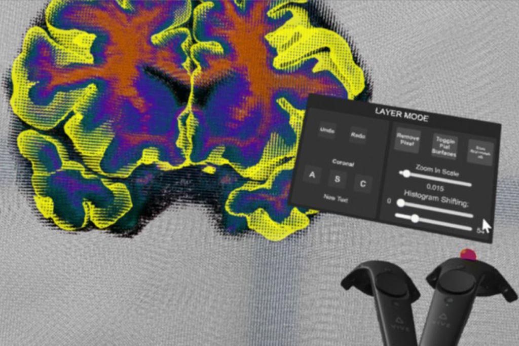
Image: The Virtual Brain Segmenter could increase the efficiency of brain scan analysis (Photo courtesy of Dominique Duncan).
Researchers at the Mark and Mary Stevens Neuroimaging and Informatics Institute at the Keck School of Medicine of University of Southern California {(USC), Los Angeles, CA, USA} have designed a virtual reality (VR) tool for correcting errors in brain scan data. The tool named Virtual Brain Segmenter (VBS) transforms a tedious step in the scientific process into an immersive experience and significantly increases the efficiency of brain scan analysis.
During automatic processing of the MRI data collected from study participants by the researchers, the segmentation step separates the brain into labeled regions to allow for close examination of the different structures. However, the automation has some drawbacks and researchers are required to manually correct the errors before proceeding with the analysis. This can be time consuming work and requires labs to hire undergraduate research assistants to correct the segmentation errors, leading to wasted time and resources spent on training.
In order to make the process more efficient and intuitive, and correct the segmentation errors with less training, the researchers launched an experimental trial to test the efficacy of VR in brain segmentation. The researchers tested 30 participants who were completely new to brain segmentation on using a MacBook to perform a minor error correction with a data cleanup program and completing the same task in VBS with a VR headset and two controllers.
They found that the participants who used VBS as compared to those using the data cleanup program finished the correction 68 seconds faster, translating into significant time savings as the task usually requires less than three minutes to be completed.
“This project is particularly exciting because not much has been done with VR in the field of neuroscience,” said Arthur Toga, provost professor and director of the neuroimaging and informatics institute. “Our main goal is to put this tool in the hands of researchers around the world. We see that as a way to continue driving discovery in the field.”
Related Links:
University of Southern California
Latest Imaging IT News
- New Google Cloud Medical Imaging Suite Makes Imaging Healthcare Data More Accessible
- Global AI in Medical Diagnostics Market to Be Driven by Demand for Image Recognition in Radiology
- AI-Based Mammography Triage Software Helps Dramatically Improve Interpretation Process
- Artificial Intelligence (AI) Program Accurately Predicts Lung Cancer Risk from CT Images
- Image Management Platform Streamlines Treatment Plans
- AI-Based Technology for Ultrasound Image Analysis Receives FDA Approval
- AI Technology for Detecting Breast Cancer Receives CE Mark Approval
- Digital Pathology Software Improves Workflow Efficiency
- Patient-Centric Portal Facilitates Direct Imaging Access
- New Workstation Supports Customer-Driven Imaging Workflow
Channels
Radiography
view channel
Routine Mammograms Could Predict Future Cardiovascular Disease in Women
Mammograms are widely used to screen for breast cancer, but they may also contain overlooked clues about cardiovascular health. Calcium deposits in the arteries of the breast signal stiffening blood vessels,... Read more
AI Detects Early Signs of Aging from Chest X-Rays
Chronological age does not always reflect how fast the body is truly aging, and current biological age tests often rely on DNA-based markers that may miss early organ-level decline. Detecting subtle, age-related... Read moreMRI
view channel
MRI Scans Reveal Signature Patterns of Brain Activity to Predict Recovery from TBI
Recovery after traumatic brain injury (TBI) varies widely, with some patients regaining full function while others are left with lasting disabilities. Prognosis is especially difficult to assess in patients... Read more
Novel Imaging Approach to Improve Treatment for Spinal Cord Injuries
Vascular dysfunction in the spinal cord contributes to multiple neurological conditions, including traumatic injuries and degenerative cervical myelopathy, where reduced blood flow can lead to progressive... Read more
AI-Assisted Model Enhances MRI Heart Scans
A cardiac MRI can reveal critical information about the heart’s function and any abnormalities, but traditional scans take 30 to 90 minutes and often suffer from poor image quality due to patient movement.... Read more
AI Model Outperforms Doctors at Identifying Patients Most At-Risk of Cardiac Arrest
Hypertrophic cardiomyopathy is one of the most common inherited heart conditions and a leading cause of sudden cardiac death in young individuals and athletes. While many patients live normal lives, some... Read moreUltrasound
view channel
Wearable Ultrasound Imaging System to Enable Real-Time Disease Monitoring
Chronic conditions such as hypertension and heart failure require close monitoring, yet today’s ultrasound imaging is largely confined to hospitals and short, episodic scans. This reactive model limits... Read more
Ultrasound Technique Visualizes Deep Blood Vessels in 3D Without Contrast Agents
Producing clear 3D images of deep blood vessels has long been difficult without relying on contrast agents, CT scans, or MRI. Standard ultrasound typically provides only 2D cross-sections, limiting clinicians’... Read moreNuclear Medicine
view channel
PET Imaging of Inflammation Predicts Recovery and Guides Therapy After Heart Attack
Acute myocardial infarction can trigger lasting heart damage, yet clinicians still lack reliable tools to identify which patients will regain function and which may develop heart failure.... Read more
Radiotheranostic Approach Detects, Kills and Reprograms Aggressive Cancers
Aggressive cancers such as osteosarcoma and glioblastoma often resist standard therapies, thrive in hostile tumor environments, and recur despite surgery, radiation, or chemotherapy. These tumors also... Read more
New Imaging Solution Improves Survival for Patients with Recurring Prostate Cancer
Detecting recurrent prostate cancer remains one of the most difficult challenges in oncology, as standard imaging methods such as bone scans and CT scans often fail to accurately locate small or early-stage tumors.... Read moreGeneral/Advanced Imaging
view channel
AI-Based Tool Accelerates Detection of Kidney Cancer
Diagnosing kidney cancer depends on computed tomography scans, often using contrast agents to reveal abnormalities in kidney structure. Tumors are not always searched for deliberately, as many scans are... Read more
New Algorithm Dramatically Speeds Up Stroke Detection Scans
When patients arrive at emergency rooms with stroke symptoms, clinicians must rapidly determine whether the cause is a blood clot or a brain bleed, as treatment decisions depend on this distinction.... Read moreImaging IT
view channel
New Google Cloud Medical Imaging Suite Makes Imaging Healthcare Data More Accessible
Medical imaging is a critical tool used to diagnose patients, and there are billions of medical images scanned globally each year. Imaging data accounts for about 90% of all healthcare data1 and, until... Read more


