Quantitative Ultrasound Helps Stage Fatty Liver
|
By MedImaging International staff writers Posted on 17 Feb 2016 |

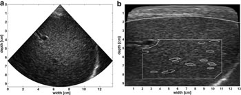
Image: Original B-Mode image next to back scan-converted B-mode image with interactively drawn fat layer contour (white dotted line); initial fixed region of interest (white rectangle); automatic segmentation within region of interest (white ellipses) (Photo courtesy of Ultrasound in Medicine & Biology).
A new study suggests that computer-aided ultrasound could be used to stage hepatic steatosis, with a high correlation to magnetic resonance imaging (MRI) spectroscopy.
Researchers at Radboud University Medical Center (Nijmegen, the Netherlands) conducted a pilot study in 14 patients on long-term home parenteral nutrition using a phased array ultrasound transducer connected to a calibrated ultrasound machine, with the radio frequency (RF) data converted into conventional B-mode images with the aid of computer software. All patients were also subjected to proton MRI spectroscopy measurement of liver fat content for reference.
The results showed that computer-aided ultrasound parameters, similar to those in a previous validation study in cows, demonstrated significant correlation with fat content as measured by MRI spectroscopy, with the most significant parameters being residual attenuation coefficient and lateral speckle size. According to the researchers, the method shows promise as a noninvasive, easy, and inexpensive alternative to invasive biopsies or expensive MRI spectroscopy techniques. The study was published in the March 2016 issue of Ultrasound in Medicine & Biology.
“Patients on home parenteral nutrition are at risk for developing liver dysfunction, which is due partly to the accumulation of lipids in the liver and may progress to end-stage liver disease with overt liver failure,” said lead author Gert Weijers, PhD, of the Radboud Medical Ultrasound Imaging Centre (MUSIC). “Therefore, a timely diagnosis with easy access to repeated assessment of the degree of liver steatosis is of great importance.”
Steatosis in the liver, when it is not due to excessive alcohol use, is most often related to insulin resistance and to metabolic syndrome, and as a result may respond to treatments originally developed for other insulin-resistant states, such as diabetes mellitus type 2. Steatosis is also the most common liver disease of high-yielding dairy cattle during early lactation, making it a suitable animal model for studying liver fat accumulation.
Related Links:
Radboud University Medical Center
Latest Ultrasound News
- Wearable Ultrasound Imaging System to Enable Real-Time Disease Monitoring
- Ultrasound Technique Visualizes Deep Blood Vessels in 3D Without Contrast Agents
- Ultrasound Probe Images Entire Organ in 4D

- Disposable Ultrasound Patch Performs Better Than Existing Devices
- Non-Invasive Ultrasound-Based Tool Accurately Detects Infant Meningitis
- Breakthrough Deep Learning Model Enhances Handheld 3D Medical Imaging
- Pain-Free Breast Imaging System Performs One Minute Cancer Scan
- Wireless Chronic Pain Management Device to Reduce Need for Painkillers and Surgery
- New Medical Ultrasound Imaging Technique Enables ICU Bedside Monitoring
- New Incision-Free Technique Halts Growth of Debilitating Brain Lesions
- AI-Powered Lung Ultrasound Outperforms Human Experts in Tuberculosis Diagnosis
- AI Identifies Heart Valve Disease from Common Imaging Test
- Novel Imaging Method Enables Early Diagnosis and Treatment Monitoring of Type 2 Diabetes
- Ultrasound-Based Microscopy Technique to Help Diagnose Small Vessel Diseases
- Smart Ultrasound-Activated Immune Cells Destroy Cancer Cells for Extended Periods
- Tiny Magnetic Robot Takes 3D Scans from Deep Within Body
Channels
Radiography
view channel
Routine Mammograms Could Predict Future Cardiovascular Disease in Women
Mammograms are widely used to screen for breast cancer, but they may also contain overlooked clues about cardiovascular health. Calcium deposits in the arteries of the breast signal stiffening blood vessels,... Read more
AI Detects Early Signs of Aging from Chest X-Rays
Chronological age does not always reflect how fast the body is truly aging, and current biological age tests often rely on DNA-based markers that may miss early organ-level decline. Detecting subtle, age-related... Read moreMRI
view channel
MRI Scans Reveal Signature Patterns of Brain Activity to Predict Recovery from TBI
Recovery after traumatic brain injury (TBI) varies widely, with some patients regaining full function while others are left with lasting disabilities. Prognosis is especially difficult to assess in patients... Read more
Novel Imaging Approach to Improve Treatment for Spinal Cord Injuries
Vascular dysfunction in the spinal cord contributes to multiple neurological conditions, including traumatic injuries and degenerative cervical myelopathy, where reduced blood flow can lead to progressive... Read more
AI-Assisted Model Enhances MRI Heart Scans
A cardiac MRI can reveal critical information about the heart’s function and any abnormalities, but traditional scans take 30 to 90 minutes and often suffer from poor image quality due to patient movement.... Read more
AI Model Outperforms Doctors at Identifying Patients Most At-Risk of Cardiac Arrest
Hypertrophic cardiomyopathy is one of the most common inherited heart conditions and a leading cause of sudden cardiac death in young individuals and athletes. While many patients live normal lives, some... Read moreNuclear Medicine
view channel
PET Imaging of Inflammation Predicts Recovery and Guides Therapy After Heart Attack
Acute myocardial infarction can trigger lasting heart damage, yet clinicians still lack reliable tools to identify which patients will regain function and which may develop heart failure.... Read more
Radiotheranostic Approach Detects, Kills and Reprograms Aggressive Cancers
Aggressive cancers such as osteosarcoma and glioblastoma often resist standard therapies, thrive in hostile tumor environments, and recur despite surgery, radiation, or chemotherapy. These tumors also... Read more
New Imaging Solution Improves Survival for Patients with Recurring Prostate Cancer
Detecting recurrent prostate cancer remains one of the most difficult challenges in oncology, as standard imaging methods such as bone scans and CT scans often fail to accurately locate small or early-stage tumors.... Read moreGeneral/Advanced Imaging
view channel
AI-Based Tool Accelerates Detection of Kidney Cancer
Diagnosing kidney cancer depends on computed tomography scans, often using contrast agents to reveal abnormalities in kidney structure. Tumors are not always searched for deliberately, as many scans are... Read more
New Algorithm Dramatically Speeds Up Stroke Detection Scans
When patients arrive at emergency rooms with stroke symptoms, clinicians must rapidly determine whether the cause is a blood clot or a brain bleed, as treatment decisions depend on this distinction.... Read moreImaging IT
view channel
New Google Cloud Medical Imaging Suite Makes Imaging Healthcare Data More Accessible
Medical imaging is a critical tool used to diagnose patients, and there are billions of medical images scanned globally each year. Imaging data accounts for about 90% of all healthcare data1 and, until... Read more
Global AI in Medical Diagnostics Market to Be Driven by Demand for Image Recognition in Radiology
The global artificial intelligence (AI) in medical diagnostics market is expanding with early disease detection being one of its key applications and image recognition becoming a compelling consumer proposition... Read moreIndustry News
view channel
GE HealthCare and NVIDIA Collaboration to Reimagine Diagnostic Imaging
GE HealthCare (Chicago, IL, USA) has entered into a collaboration with NVIDIA (Santa Clara, CA, USA), expanding the existing relationship between the two companies to focus on pioneering innovation in... Read more
Patient-Specific 3D-Printed Phantoms Transform CT Imaging
New research has highlighted how anatomically precise, patient-specific 3D-printed phantoms are proving to be scalable, cost-effective, and efficient tools in the development of new CT scan algorithms... Read more
Siemens and Sectra Collaborate on Enhancing Radiology Workflows
Siemens Healthineers (Forchheim, Germany) and Sectra (Linköping, Sweden) have entered into a collaboration aimed at enhancing radiologists' diagnostic capabilities and, in turn, improving patient care... Read more


