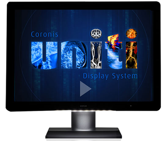
First Diagnostic Display Cleared for Viewing All Types of Breast Imaging
|
By MedImaging International staff writers Posted on 04 Nov 2015 |
A healthcare imaging specialist vendor has announce that one of the most prominent diagnostic displays it produces has been clinically validated for use for both Picture Archiving and Communication System (PACS) and multimodality breast imaging.
The US Food and Drug Administration (FDA; Silver Spring, MD USA) indicated the display for use in breast Magnetic Resonance Imaging (MRI), Computed Tomography (CT) and ultrasound, breast ultrasound, vascular and gynecological ultrasound on a single display, in grayscale, color, 2D or 3D, static or dynamic. The display had been previously cleared for viewing of mammography images, breast tomosynthesis, and PACS images.
The Coronis Uniti display was announced by Barco (Kortrijk, Belgium) and can used to access any image on a single workstation, saving time, and providing workflow and clinical benefits. The ability to run side-by-side comparisons and fuse images such as those from breast ultrasound and conventional mammography in women with dense breasts, has resulted in better detection of early breast cancer. The display can automatically set the correct color and grayscale settings for every image modality.
Coronis Uniti also enables radiologists to accurately visualize moving images in a 3D stack, ensuring efficient diagnosis, and a rapid workflow to prevent loss of detail while scrolling. Barco also found a way to counteract motion blur when reviewing image sequences.
Lynda Domogalla, Barco’s VP product marketing for Healthcare, said, “With Coronis Uniti, we wanted to break through the technical boundaries of multimodality integration and deliver the first unified workflow for radiologists in order to improve reading productivity as well health outcomes. We have succeeded in developing a ‘one size fits all’ diagnostic display—which defines a new bar for clinically-focused display solutions. To maximize the diagnostic value of color breast images, we developed new technologies to calibrate and maintain the consistency of these color images, and we are leading in the definition of a Color Standard Display Function (CSDF) to ensure the accuracy and consistency of color images on diagnostic displays.”
Related Links:
Barco
Read the full article by registering today, it's FREE! 

Register now for FREE to MedImaging.net and get access to news and events that shape the world of Radiology. 
- Free digital version edition of Medical Imaging International sent by email on regular basis
- Free print version of Medical Imaging International magazine (available only outside USA and Canada).
- Free and unlimited access to back issues of Medical Imaging International in digital format
- Free Medical Imaging International Newsletter sent every week containing the latest news
- Free breaking news sent via email
- Free access to Events Calendar
- Free access to LinkXpress new product services
- REGISTRATION IS FREE AND EASY!
Sign in: Registered website members
Sign in: Registered magazine subscribers
Latest Imaging IT News
- New Google Cloud Medical Imaging Suite Makes Imaging Healthcare Data More Accessible
- Global AI in Medical Diagnostics Market to Be Driven by Demand for Image Recognition in Radiology
- AI-Based Mammography Triage Software Helps Dramatically Improve Interpretation Process
- Artificial Intelligence (AI) Program Accurately Predicts Lung Cancer Risk from CT Images
- Image Management Platform Streamlines Treatment Plans
- AI-Based Technology for Ultrasound Image Analysis Receives FDA Approval
- AI Technology for Detecting Breast Cancer Receives CE Mark Approval
- Digital Pathology Software Improves Workflow Efficiency
- Patient-Centric Portal Facilitates Direct Imaging Access
- New Workstation Supports Customer-Driven Imaging Workflow
Channels
Radiography
view channel
World's Largest Class Single Crystal Diamond Radiation Detector Opens New Possibilities for Diagnostic Imaging
Diamonds possess ideal physical properties for radiation detection, such as exceptional thermal and chemical stability along with a quick response time. Made of carbon with an atomic number of six, diamonds... Read more
AI-Powered Imaging Technique Shows Promise in Evaluating Patients for PCI
Percutaneous coronary intervention (PCI), also known as coronary angioplasty, is a minimally invasive procedure where small metal tubes called stents are inserted into partially blocked coronary arteries... Read moreMRI
view channel
AI Tool Predicts Relapse of Pediatric Brain Cancer from Brain MRI Scans
Many pediatric gliomas are treatable with surgery alone, but relapses can be catastrophic. Predicting which patients are at risk for recurrence remains challenging, leading to frequent follow-ups with... Read more
AI Tool Tracks Effectiveness of Multiple Sclerosis Treatments Using Brain MRI Scans
Multiple sclerosis (MS) is a condition in which the immune system attacks the brain and spinal cord, leading to impairments in movement, sensation, and cognition. Magnetic Resonance Imaging (MRI) markers... Read more
Ultra-Powerful MRI Scans Enable Life-Changing Surgery in Treatment-Resistant Epileptic Patients
Approximately 360,000 individuals in the UK suffer from focal epilepsy, a condition in which seizures spread from one part of the brain. Around a third of these patients experience persistent seizures... Read moreUltrasound
view channel.jpeg)
AI-Powered Lung Ultrasound Outperforms Human Experts in Tuberculosis Diagnosis
Despite global declines in tuberculosis (TB) rates in previous years, the incidence of TB rose by 4.6% from 2020 to 2023. Early screening and rapid diagnosis are essential elements of the World Health... Read more
AI Identifies Heart Valve Disease from Common Imaging Test
Tricuspid regurgitation is a condition where the heart's tricuspid valve does not close completely during contraction, leading to backward blood flow, which can result in heart failure. A new artificial... Read moreNuclear Medicine
view channel
Novel Radiolabeled Antibody Improves Diagnosis and Treatment of Solid Tumors
Interleukin-13 receptor α-2 (IL13Rα2) is a cell surface receptor commonly found in solid tumors such as glioblastoma, melanoma, and breast cancer. It is minimally expressed in normal tissues, making it... Read more
Novel PET Imaging Approach Offers Never-Before-Seen View of Neuroinflammation
COX-2, an enzyme that plays a key role in brain inflammation, can be significantly upregulated by inflammatory stimuli and neuroexcitation. Researchers suggest that COX-2 density in the brain could serve... Read moreGeneral/Advanced Imaging
view channel
AI-Powered Imaging System Improves Lung Cancer Diagnosis
Given the need to detect lung cancer at earlier stages, there is an increasing need for a definitive diagnostic pathway for patients with suspicious pulmonary nodules. However, obtaining tissue samples... Read more
AI Model Significantly Enhances Low-Dose CT Capabilities
Lung cancer remains one of the most challenging diseases, making early diagnosis vital for effective treatment. Fortunately, advancements in artificial intelligence (AI) are revolutionizing lung cancer... Read moreIndustry News
view channel
GE HealthCare and NVIDIA Collaboration to Reimagine Diagnostic Imaging
GE HealthCare (Chicago, IL, USA) has entered into a collaboration with NVIDIA (Santa Clara, CA, USA), expanding the existing relationship between the two companies to focus on pioneering innovation in... Read more
Patient-Specific 3D-Printed Phantoms Transform CT Imaging
New research has highlighted how anatomically precise, patient-specific 3D-printed phantoms are proving to be scalable, cost-effective, and efficient tools in the development of new CT scan algorithms... Read more
Siemens and Sectra Collaborate on Enhancing Radiology Workflows
Siemens Healthineers (Forchheim, Germany) and Sectra (Linköping, Sweden) have entered into a collaboration aimed at enhancing radiologists' diagnostic capabilities and, in turn, improving patient care... Read more


