Bone Suppression Software Used to Optimize Diagnostic Capability of X-Ray Systems
|
By MedImaging International staff writers Posted on 18 Aug 2014 |
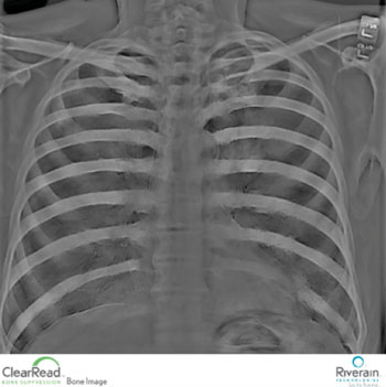
Clinicians are gathering important information from the most routine imaging exam, the chest X-ray, by using advanced software that enhances X-ray images captured by the equipment they already have or are in the process of buying. New bone suppression technology helps radiologists identify lung nodules and other serious medical conditions by converting a traditional chest X-ray into a soft tissue image without the ribs and clavicle bones.
These structures typically obscure abnormalities. There are no further testing or radiation exposure for patients, and no need for imaging units or hardware for the hospital to buy or house to achieve the soft tissue image.
Riverain Technologies (Dayton, OH, USA) revenues increased 34% year-over-year through the first half of 2014 based on strong adoption of the company’s ClearRead software solutions. This was particularly true among academic medical centers and community and Veterans Administration (VA) hospitals throughout the US Midwest. “Most of the growth in adoption is being driven by radiologists’ recognition that bone suppression is very useful and there’s little to no downside to it,” said Heber MacMahon, MB, BCh, professor of radiology and director of thoracic imaging at the University of Chicago Medical Center (IL, USA).
In the past, no-bone, soft tissue images were only made possible by exposing patients to higher radiation levels using costly imaging equipment. The enhancements in clarity and performance of the X-ray are advantageous for patients because that X-ray is the most readily available and widely used imaging modality. Many lung tumors are first targeted on X-ray images taken for other reasons, such as to diagnose other medical conditions, including heart problems, broken ribs, collapsed lung, pneumonia, and emphysema.
“It’s nice to get more out of what we deliver,” said Ella Kazerooni, MD, past president of the Society of Thoracic Radiology, and professor and director, division cardiothoracic radiology, and associate chair for clinical affairs at the University of Michigan (U-M) Health System (Ann Arbor, USA). “The software is easy to install, easy to train to, and, frankly it provides immediate clinical value.”
The software is sold directly to medical centers, to enhance X-ray images captured by the machines they already have, and by major manufacturers including Siemens Healthcare (Erlangen, Germany) and Philips Healthcare (Best, The Netherlands) as a value-added extension to new X-ray equipment.
University of Chicago (IL, USA) bought ClearRead software after serving as a research site and validating the effectiveness of Riverain’s ClearRead bone suppression and computer-aided diagnosis (CAD) software, ClearRead +Detect, in improving the diagnostic potential of traditional X-ray images. “As long as we’re going to continue to do millions of chest X-rays around the world, with this software we’re going to pick up more cancers sooner and we’re going to miss less cancers,” said Dr. MacMahon, who is a member of Riverain’s medical advisory board.
In addition to purchasing ClearRead bone suppression and ClearRead + Detect software, which marks suspected lung nodules on an X-ray image, U-M Health System physicians are planning on purchasing of ClearRead +Confirm software, based on a clinical evaluation of the software.
ClearRead +Confirm identifies and highlights lines and tubes on portable chest X-ray images, reducing reading time by approximately 19% without compromising radiologist accuracy or confidence. “Referring physicians working in our intensive care units can see lines and tubes much more clearly without having to make adjustments to each image, line by line and case by case,” Dr. Kazerooni said. “They’re bypassing the standard X-ray image and going directly to the Riverain-processed images. This speaks to how highly they regard the software.”
The benefit of ClearRead +Confirm for patients, Dr. Kazerooni added, is that “It gives us greater confidence about the devices we’re putting in place so that we’re sure they’re functioning optimally.”
Riverain Technologies’ ClearRead software enhances the expert skills of radiologists to improve patient outcomes using standard chest X-ray, without additional radiation dose or procedures for patients. Riverain ClearRead software solutions include ClearRead bone suppression, +Detect, +Confirm and +Compare, which reveals density alterations between current and earlier chest X-ray images.
Related Links:
Riverain Technologies
Read the full article by registering today, it's FREE! 

Register now for FREE to MedImaging.net and get access to news and events that shape the world of Radiology. 
- Free digital version edition of Medical Imaging International sent by email on regular basis
- Free print version of Medical Imaging International magazine (available only outside USA and Canada).
- Free and unlimited access to back issues of Medical Imaging International in digital format
- Free Medical Imaging International Newsletter sent every week containing the latest news
- Free breaking news sent via email
- Free access to Events Calendar
- Free access to LinkXpress new product services
- REGISTRATION IS FREE AND EASY!
Sign in: Registered website members
Sign in: Registered magazine subscribers
Latest Imaging IT News
- New Google Cloud Medical Imaging Suite Makes Imaging Healthcare Data More Accessible
- Global AI in Medical Diagnostics Market to Be Driven by Demand for Image Recognition in Radiology
- AI-Based Mammography Triage Software Helps Dramatically Improve Interpretation Process
- Artificial Intelligence (AI) Program Accurately Predicts Lung Cancer Risk from CT Images
- Image Management Platform Streamlines Treatment Plans
- AI-Based Technology for Ultrasound Image Analysis Receives FDA Approval
- AI Technology for Detecting Breast Cancer Receives CE Mark Approval
- Digital Pathology Software Improves Workflow Efficiency
- Patient-Centric Portal Facilitates Direct Imaging Access
- New Workstation Supports Customer-Driven Imaging Workflow
Channels
Radiography
view channel
World's Largest Class Single Crystal Diamond Radiation Detector Opens New Possibilities for Diagnostic Imaging
Diamonds possess ideal physical properties for radiation detection, such as exceptional thermal and chemical stability along with a quick response time. Made of carbon with an atomic number of six, diamonds... Read more
AI-Powered Imaging Technique Shows Promise in Evaluating Patients for PCI
Percutaneous coronary intervention (PCI), also known as coronary angioplasty, is a minimally invasive procedure where small metal tubes called stents are inserted into partially blocked coronary arteries... Read moreMRI
view channel
AI Tool Tracks Effectiveness of Multiple Sclerosis Treatments Using Brain MRI Scans
Multiple sclerosis (MS) is a condition in which the immune system attacks the brain and spinal cord, leading to impairments in movement, sensation, and cognition. Magnetic Resonance Imaging (MRI) markers... Read more
Ultra-Powerful MRI Scans Enable Life-Changing Surgery in Treatment-Resistant Epileptic Patients
Approximately 360,000 individuals in the UK suffer from focal epilepsy, a condition in which seizures spread from one part of the brain. Around a third of these patients experience persistent seizures... Read more
AI-Powered MRI Technology Improves Parkinson’s Diagnoses
Current research shows that the accuracy of diagnosing Parkinson’s disease typically ranges from 55% to 78% within the first five years of assessment. This is partly due to the similarities shared by Parkinson’s... Read more
Biparametric MRI Combined with AI Enhances Detection of Clinically Significant Prostate Cancer
Artificial intelligence (AI) technologies are transforming the way medical images are analyzed, offering unprecedented capabilities in quantitatively extracting features that go beyond traditional visual... Read moreUltrasound
view channel.jpeg)
AI-Powered Lung Ultrasound Outperforms Human Experts in Tuberculosis Diagnosis
Despite global declines in tuberculosis (TB) rates in previous years, the incidence of TB rose by 4.6% from 2020 to 2023. Early screening and rapid diagnosis are essential elements of the World Health... Read more
AI Identifies Heart Valve Disease from Common Imaging Test
Tricuspid regurgitation is a condition where the heart's tricuspid valve does not close completely during contraction, leading to backward blood flow, which can result in heart failure. A new artificial... Read moreNuclear Medicine
view channel
Novel Radiolabeled Antibody Improves Diagnosis and Treatment of Solid Tumors
Interleukin-13 receptor α-2 (IL13Rα2) is a cell surface receptor commonly found in solid tumors such as glioblastoma, melanoma, and breast cancer. It is minimally expressed in normal tissues, making it... Read more
Novel PET Imaging Approach Offers Never-Before-Seen View of Neuroinflammation
COX-2, an enzyme that plays a key role in brain inflammation, can be significantly upregulated by inflammatory stimuli and neuroexcitation. Researchers suggest that COX-2 density in the brain could serve... Read moreGeneral/Advanced Imaging
view channel
AI-Powered Imaging System Improves Lung Cancer Diagnosis
Given the need to detect lung cancer at earlier stages, there is an increasing need for a definitive diagnostic pathway for patients with suspicious pulmonary nodules. However, obtaining tissue samples... Read more
AI Model Significantly Enhances Low-Dose CT Capabilities
Lung cancer remains one of the most challenging diseases, making early diagnosis vital for effective treatment. Fortunately, advancements in artificial intelligence (AI) are revolutionizing lung cancer... Read moreIndustry News
view channel
GE HealthCare and NVIDIA Collaboration to Reimagine Diagnostic Imaging
GE HealthCare (Chicago, IL, USA) has entered into a collaboration with NVIDIA (Santa Clara, CA, USA), expanding the existing relationship between the two companies to focus on pioneering innovation in... Read more
Patient-Specific 3D-Printed Phantoms Transform CT Imaging
New research has highlighted how anatomically precise, patient-specific 3D-printed phantoms are proving to be scalable, cost-effective, and efficient tools in the development of new CT scan algorithms... Read more
Siemens and Sectra Collaborate on Enhancing Radiology Workflows
Siemens Healthineers (Forchheim, Germany) and Sectra (Linköping, Sweden) have entered into a collaboration aimed at enhancing radiologists' diagnostic capabilities and, in turn, improving patient care... Read more


