Mammography Screening Intervals May Affect Breast Cancer Prognosis
|
By MedImaging International staff writers Posted on 02 Jan 2014 |

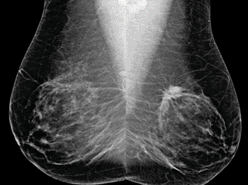
Image: Bilateral mediolateral oblique (MLO) views from screening mammography in a 53-year-old woman (Photo courtesy of RSNA).

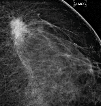
Image: Spot magnification view demonstrates an irregular spiculated mass with associated calcifications in the upper outer left breast. Ultrasound biopsy revealed invasive ductal carcinoma and DCIS (Photo courtesy of RSNA).

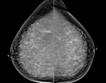
Image: Screening mammogram in a 63-year-old woman demonstrated calcifications in the upper outer left breast. Bilateral craniocaudal (CC) views shown (Photo courtesy of RSNA).
In a study of screening mammography-detected breast tumors, patients who had more frequent screening mammography exams had a considerably lower rate of lymph node positivity as compared to women who had longer intervals between screening-mammography exams.
The study’s findings were presented December 3, 2013, at the annual meeting of the Radiological Society of North America (RSNA), held December 2013 in Chicago (IL, USA). In its earliest stages, breast cancer is confined to the breast and can be treated by surgically removing the cancer cells. Breast cancer cells may metastasize to the lymph nodes as the disease progresses and then to other areas of the body.
“On its pathway to other places in the body, the first place breast cancer typically drains into before metastasizing is the lymph nodes,” said Lilian Wang, MD, assistant professor of radiology at Northwestern University/Feinberg School of Medicine (Chicago, IL, USA). “When breast cancer has spread into the lymph nodes, the patient is often treated both locally and systemically, with either hormone therapy, chemotherapy, trastuzumab, or some combination of these therapies.”
Healthcare organizations, such as RSNA and the American Cancer Society (ACS), have recommended yearly screening with mammography for women beginning at age 40. However, in 2009, the United States Preventive Services Task Force (USPSTF) announced a controversial new recommendation for biennial screening for women between the ages of 50 and 74. “Our study looks at what would happen if the revised guidelines issued by USPSTF were followed by women,” Dr. Wang said.
Conducted at Northwestern Memorial Hospital (Chicago, IL, USA), the retrospective study, included 332 women with breast cancer identified by screening mammography between 2007 and 2010. The women were split into one of three groups, based on the length of time between their screening mammography exams: less than 1.5 years, 1.5 to three years, and more than three years. There were 207, 73, and 52 patients in each category, respectively.
The researchers determined, controlling for age, breast density, high-risk status, and a family history of breast cancer, that women in the less than 1.5-year interval group had the lowest lymph node positivity rate at 8.7%. The rate of lymph node involvement was substantially higher in the 1.5- to three-year and over three-year interval groups at 20.5% and 15.4%, respectively.
“Our study shows that screening mammography performed at an interval of less than 1.5 years reduces the rate of lymph node positivity, thereby improving patient prognosis,” Dr. Wang concluded. “We should be following the guidelines of the American Cancer Society and other organizations, recommending that women undergo annual screening mammography beginning at age 40.”
Related Links:
Northwestern University/Feinberg School of Medicine
Latest Radiography News
- Machine Learning Algorithm Identifies Cardiovascular Risk from Routine Bone Density Scans
- AI Improves Early Detection of Interval Breast Cancers
- World's Largest Class Single Crystal Diamond Radiation Detector Opens New Possibilities for Diagnostic Imaging
- AI-Powered Imaging Technique Shows Promise in Evaluating Patients for PCI
- Higher Chest X-Ray Usage Catches Lung Cancer Earlier and Improves Survival
- AI-Powered Mammograms Predict Cardiovascular Risk
- Generative AI Model Significantly Reduces Chest X-Ray Reading Time
- AI-Powered Mammography Screening Boosts Cancer Detection in Single-Reader Settings
- Photon Counting Detectors Promise Fast Color X-Ray Images
- AI Can Flag Mammograms for Supplemental MRI
- 3D CT Imaging from Single X-Ray Projection Reduces Radiation Exposure
- AI Method Accurately Predicts Breast Cancer Risk by Analyzing Multiple Mammograms
- Printable Organic X-Ray Sensors Could Transform Treatment for Cancer Patients
- Highly Sensitive, Foldable Detector to Make X-Rays Safer
- Novel Breast Cancer Screening Technology Could Offer Superior Alternative to Mammogram
- Artificial Intelligence Accurately Predicts Breast Cancer Years Before Diagnosis
Channels
MRI
view channel
Cutting-Edge MRI Technology to Revolutionize Diagnosis of Common Heart Problem
Aortic stenosis is a common and potentially life-threatening heart condition. It occurs when the aortic valve, which regulates blood flow from the heart to the rest of the body, becomes stiff and narrow.... Read more
New MRI Technique Reveals True Heart Age to Prevent Attacks and Strokes
Heart disease remains one of the leading causes of death worldwide. Individuals with conditions such as diabetes or obesity often experience accelerated aging of their hearts, sometimes by decades.... Read more
AI Tool Predicts Relapse of Pediatric Brain Cancer from Brain MRI Scans
Many pediatric gliomas are treatable with surgery alone, but relapses can be catastrophic. Predicting which patients are at risk for recurrence remains challenging, leading to frequent follow-ups with... Read more
AI Tool Tracks Effectiveness of Multiple Sclerosis Treatments Using Brain MRI Scans
Multiple sclerosis (MS) is a condition in which the immune system attacks the brain and spinal cord, leading to impairments in movement, sensation, and cognition. Magnetic Resonance Imaging (MRI) markers... Read moreUltrasound
view channel.jpeg)
AI-Powered Lung Ultrasound Outperforms Human Experts in Tuberculosis Diagnosis
Despite global declines in tuberculosis (TB) rates in previous years, the incidence of TB rose by 4.6% from 2020 to 2023. Early screening and rapid diagnosis are essential elements of the World Health... Read more
AI Identifies Heart Valve Disease from Common Imaging Test
Tricuspid regurgitation is a condition where the heart's tricuspid valve does not close completely during contraction, leading to backward blood flow, which can result in heart failure. A new artificial... Read moreNuclear Medicine
view channel
Novel Radiolabeled Antibody Improves Diagnosis and Treatment of Solid Tumors
Interleukin-13 receptor α-2 (IL13Rα2) is a cell surface receptor commonly found in solid tumors such as glioblastoma, melanoma, and breast cancer. It is minimally expressed in normal tissues, making it... Read more
Novel PET Imaging Approach Offers Never-Before-Seen View of Neuroinflammation
COX-2, an enzyme that plays a key role in brain inflammation, can be significantly upregulated by inflammatory stimuli and neuroexcitation. Researchers suggest that COX-2 density in the brain could serve... Read moreGeneral/Advanced Imaging
view channel
AI-Based CT Scan Analysis Predicts Early-Stage Kidney Damage Due to Cancer Treatments
Radioligand therapy, a form of targeted nuclear medicine, has recently gained attention for its potential in treating specific types of tumors. However, one of the potential side effects of this therapy... Read more
CT-Based Deep Learning-Driven Tool to Enhance Liver Cancer Diagnosis
Medical imaging, such as computed tomography (CT) scans, plays a crucial role in oncology, offering essential data for cancer detection, treatment planning, and monitoring of response to therapies.... Read moreImaging IT
view channel
New Google Cloud Medical Imaging Suite Makes Imaging Healthcare Data More Accessible
Medical imaging is a critical tool used to diagnose patients, and there are billions of medical images scanned globally each year. Imaging data accounts for about 90% of all healthcare data1 and, until... Read more
Global AI in Medical Diagnostics Market to Be Driven by Demand for Image Recognition in Radiology
The global artificial intelligence (AI) in medical diagnostics market is expanding with early disease detection being one of its key applications and image recognition becoming a compelling consumer proposition... Read moreIndustry News
view channel
GE HealthCare and NVIDIA Collaboration to Reimagine Diagnostic Imaging
GE HealthCare (Chicago, IL, USA) has entered into a collaboration with NVIDIA (Santa Clara, CA, USA), expanding the existing relationship between the two companies to focus on pioneering innovation in... Read more
Patient-Specific 3D-Printed Phantoms Transform CT Imaging
New research has highlighted how anatomically precise, patient-specific 3D-printed phantoms are proving to be scalable, cost-effective, and efficient tools in the development of new CT scan algorithms... Read more
Siemens and Sectra Collaborate on Enhancing Radiology Workflows
Siemens Healthineers (Forchheim, Germany) and Sectra (Linköping, Sweden) have entered into a collaboration aimed at enhancing radiologists' diagnostic capabilities and, in turn, improving patient care... Read more


