New Software Designed to Streamline Nuclear Medicine Workflow
|
By MedImaging International staff writers Posted on 08 Aug 2013 |
New nuclear medicine software has been developed to fully integrate and streamline a hospital’s nuclear medicine department workflow and enhance interdepartmental communications.
Specialized functionalities include processing, reading, and reporting of all nuclear medicine, single photon emission tomography (SPECT), positron emission tomography (PET), and multimodality images.
Agfa HealthCare (Mortsel, Belgium) has signed an agreement with Ghent University Hospital (UZG; Belgium) to implement its Impaxfor Nuclear Medicine solution, including the Oasis for Impax dedicated nuclear medicine workstation package, within the hospital. The system is expected to be operational by the end of 2013 and will complement UZG’s Impax radiology information system/picture archiving and communication system (RIS/PACS).
UZG is one of the largest hospitals in the Flanders region of Belgium. Every medical specialty for regular and intensive care is represented in this 1,000-plus bed hospital, which treats approximately 75,000 inpatients and 400,000 outpatients annually. Its goal is to use treatment, education, and research to improve the lives of patients. Enhancing decision-making and analysis for diagnosis is part of this aim.
Impax for Nuclear Medicine is built on Agfa HealthCare’s Impax 6 RIS/PACS/Reporting workflow platform for image and data management. Designed to meet the unique needs of the specialty, it allows hospitals to fully integrate the nuclear medicine department’s workflow and to optimize interdepartmental communications. It comprises specialized functionalities to process, read, and report all nuclear medicine, SPECT, PET, and multimodality images. User-friendly features include guided workflows that incorporate applications, data, and image processing within one unified image management and reporting application.
Impax for Nuclear Medicine comprises the Oasis for Impax workstation, developed by Agfa HealthCare in partnership with Segami Corp. (Columbia, MD, USA), which brings an integrated, fully featured nuclear medicine image processing and viewing package to the Impax workstation. Clinicians no longer have to switch between workstations, or manually synchronize patient study lists.
“Impax for Nuclear Medicine will allow us to easily consult and compare prior exams and data,” noted Prof. Ingeborg Goethals, head of the nuclear medicine department. “The solution is already being used in several sites in Europe, the Middle-East, and North America, so we were confident that it would also work well for us.”
Related Links:
Agfa HealthCare
Ghent University Hospital
Segami
Latest Imaging IT News
- New Google Cloud Medical Imaging Suite Makes Imaging Healthcare Data More Accessible
- Global AI in Medical Diagnostics Market to Be Driven by Demand for Image Recognition in Radiology
- AI-Based Mammography Triage Software Helps Dramatically Improve Interpretation Process
- Artificial Intelligence (AI) Program Accurately Predicts Lung Cancer Risk from CT Images
- Image Management Platform Streamlines Treatment Plans
- AI-Based Technology for Ultrasound Image Analysis Receives FDA Approval
- AI Technology for Detecting Breast Cancer Receives CE Mark Approval
- Digital Pathology Software Improves Workflow Efficiency
- Patient-Centric Portal Facilitates Direct Imaging Access
- New Workstation Supports Customer-Driven Imaging Workflow
Channels
Radiography
view channel
AI Improves Early Detection of Interval Breast Cancers
Interval breast cancers, which occur between routine screenings, are easier to treat when detected earlier. Early detection can reduce the need for aggressive treatments and improve the chances of better outcomes.... Read more
World's Largest Class Single Crystal Diamond Radiation Detector Opens New Possibilities for Diagnostic Imaging
Diamonds possess ideal physical properties for radiation detection, such as exceptional thermal and chemical stability along with a quick response time. Made of carbon with an atomic number of six, diamonds... Read moreMRI
view channel
New MRI Technique Reveals True Heart Age to Prevent Attacks and Strokes
Heart disease remains one of the leading causes of death worldwide. Individuals with conditions such as diabetes or obesity often experience accelerated aging of their hearts, sometimes by decades.... Read more
AI Tool Predicts Relapse of Pediatric Brain Cancer from Brain MRI Scans
Many pediatric gliomas are treatable with surgery alone, but relapses can be catastrophic. Predicting which patients are at risk for recurrence remains challenging, leading to frequent follow-ups with... Read more
AI Tool Tracks Effectiveness of Multiple Sclerosis Treatments Using Brain MRI Scans
Multiple sclerosis (MS) is a condition in which the immune system attacks the brain and spinal cord, leading to impairments in movement, sensation, and cognition. Magnetic Resonance Imaging (MRI) markers... Read more
Ultra-Powerful MRI Scans Enable Life-Changing Surgery in Treatment-Resistant Epileptic Patients
Approximately 360,000 individuals in the UK suffer from focal epilepsy, a condition in which seizures spread from one part of the brain. Around a third of these patients experience persistent seizures... Read moreUltrasound
view channel.jpeg)
AI-Powered Lung Ultrasound Outperforms Human Experts in Tuberculosis Diagnosis
Despite global declines in tuberculosis (TB) rates in previous years, the incidence of TB rose by 4.6% from 2020 to 2023. Early screening and rapid diagnosis are essential elements of the World Health... Read more
AI Identifies Heart Valve Disease from Common Imaging Test
Tricuspid regurgitation is a condition where the heart's tricuspid valve does not close completely during contraction, leading to backward blood flow, which can result in heart failure. A new artificial... Read moreNuclear Medicine
view channel
Novel Radiolabeled Antibody Improves Diagnosis and Treatment of Solid Tumors
Interleukin-13 receptor α-2 (IL13Rα2) is a cell surface receptor commonly found in solid tumors such as glioblastoma, melanoma, and breast cancer. It is minimally expressed in normal tissues, making it... Read more
Novel PET Imaging Approach Offers Never-Before-Seen View of Neuroinflammation
COX-2, an enzyme that plays a key role in brain inflammation, can be significantly upregulated by inflammatory stimuli and neuroexcitation. Researchers suggest that COX-2 density in the brain could serve... Read moreGeneral/Advanced Imaging
view channel
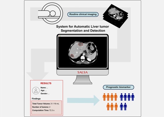
CT-Based Deep Learning-Driven Tool to Enhance Liver Cancer Diagnosis
Medical imaging, such as computed tomography (CT) scans, plays a crucial role in oncology, offering essential data for cancer detection, treatment planning, and monitoring of response to therapies.... Read more
AI-Powered Imaging System Improves Lung Cancer Diagnosis
Given the need to detect lung cancer at earlier stages, there is an increasing need for a definitive diagnostic pathway for patients with suspicious pulmonary nodules. However, obtaining tissue samples... Read moreIndustry News
view channel
GE HealthCare and NVIDIA Collaboration to Reimagine Diagnostic Imaging
GE HealthCare (Chicago, IL, USA) has entered into a collaboration with NVIDIA (Santa Clara, CA, USA), expanding the existing relationship between the two companies to focus on pioneering innovation in... Read more
Patient-Specific 3D-Printed Phantoms Transform CT Imaging
New research has highlighted how anatomically precise, patient-specific 3D-printed phantoms are proving to be scalable, cost-effective, and efficient tools in the development of new CT scan algorithms... Read more
Siemens and Sectra Collaborate on Enhancing Radiology Workflows
Siemens Healthineers (Forchheim, Germany) and Sectra (Linköping, Sweden) have entered into a collaboration aimed at enhancing radiologists' diagnostic capabilities and, in turn, improving patient care... Read more


