Computational Imaging Technique Creates 3D Model of Mammal Lung Image
|
By MedImaging International staff writers Posted on 16 Oct 2012 |
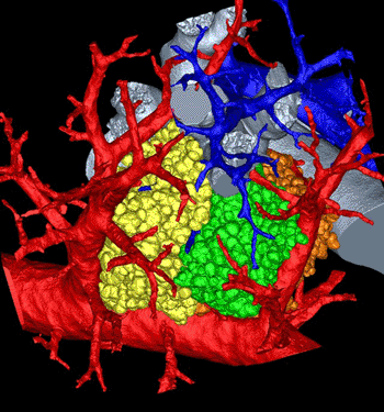
Image: Lung maze modeled in 3D (Photo courtesy of Dragos Vasilescu, University of Iowa and the University of British Columbia).
Scientists are trying to determine more precisely what occurs in the lung’s minuscule, convoluted network of corridors. To accomplish this goal, investigators have created the most detailed, three-dimensional (3D) rendering of the ending of all the pathways in the lung, called the pulmonary acinus.
The computerized model, derived from mice, authentically imitates the curves in this area, including the direction, length, and angles of the respiratory branches that lead to the air sacs called alveoli. “The imaging and image analysis methods described here provide for branch morphometry at the acinar level that has not been available previously,” the researchers, from the University of Iowa (UI; Iowa City, USA), wrote in their article, published in October 2012 in the online early edition of the Proceedings of the National Academy of Sciences of the United States of America (PNAS).
The model is significant because it can help scientists understand where and how lung diseases emerge as well as the role the pulmonary acinus plays in drug delivery, such as those typically administered with inhalers. “These methods allow us to understand where in the lung periphery disease begins and how it progresses,” said Dr. Eric Hoffman, professor in the departments of radiology, medicine, and biomedical engineering at the UI and corresponding author of the article. “How do gases and inhaled substances get there and do they accumulate in one or another acinus? How do they swirl around and clear out? We just don’t have a complete understanding how that happens.”
For instance, Dr. Hoffman reported that the model could be utilized to determine how smoking-induced emphysema begins. “It has been hypothesized recently that it begins with the loss of peripheral airways rather than the lung air sacs,” he said, mentioning ongoing research by Dr. James Hogg at the University of British Columbia (Vancouver, BC, Canada), who was not involved in this study. It also could provide insights and lead to more effective treatment of chronic obstructive pulmonary disease, which causes irreversible damage to the lung, noted Dr. Dragos Vasilescu, first author on the paper who based his thesis on the research while a graduate student at the UI.
The best, up to now, that lung anatomy specialists such as study co-corresponding author Dr. Ewald Weibel, professor emeritus of anatomy at the University of Bern (Switzerland), could do to study specific areas of a lung was to make measurements in two dimensions or create 3D casts of a lung’s air spaces. The techniques, while offering the first clues into a lung’s composition workings, had their limitations. For instance, they did not directly replicate a lung’s structure in real life, and they could not determine how various parts act together as a whole. However, developments in imaging and computer science have enabled researchers to better study how gases and other inhaled substances act in the lung’s farthest recesses.
In this study, the investigators worked with 22 pulmonary acini taken from young and old mice. They then “reconstructed” the acini based on microcomputed tomography (CT) imaging of scanned lungs in mice and extracted from them. The extracted lungs were preserved in a way that kept the anatomy intact--including the tiny air spaces required for effective imaging. From that, the researchers were able to measure an acinus, estimate the number of acini for each mouse lung, and count the alveoli and measure their surface area.
The mouse lung, in its structure and function, is remarkably similar to the human lung. That means researchers can alter the genetics of a mouse and see how those changes affect the peripheral structure of the lung and its performance. The researchers discovered that mouse alveoli increase in number a long past the two weeks that at least one earlier study had indicated. Dr. Hoffman added that more research is required to determine whether if humans also increase the number of air sacs after a specific, predetermined age.
The researcher’s next objective is to utilize the mouse model to more precisely determine how gases interact with the bloodstream within the acini and the alveoli. “Our imaging and image-analysis methodologies enable new ways to investigate the lung’s structure and can now be used to further investigate the normal healthy-lung anatomy in humans and be used to visualize and assess the pathological changes in animal models of specific structural diseases,” said Dr. Vasilescu, who is a postdoctoral research fellow at the University of British Columbia.
Related Links:
University of Iowa
University of Bern
Latest Imaging IT News
- New Google Cloud Medical Imaging Suite Makes Imaging Healthcare Data More Accessible
- Global AI in Medical Diagnostics Market to Be Driven by Demand for Image Recognition in Radiology
- AI-Based Mammography Triage Software Helps Dramatically Improve Interpretation Process
- Artificial Intelligence (AI) Program Accurately Predicts Lung Cancer Risk from CT Images
- Image Management Platform Streamlines Treatment Plans
- AI-Based Technology for Ultrasound Image Analysis Receives FDA Approval
- AI Technology for Detecting Breast Cancer Receives CE Mark Approval
- Digital Pathology Software Improves Workflow Efficiency
- Patient-Centric Portal Facilitates Direct Imaging Access
- New Workstation Supports Customer-Driven Imaging Workflow
Channels
Radiography
view channel
AI Radiology Tool Identifies Life-Threatening Conditions in Milliseconds
Radiology is emerging as one of healthcare’s most pressing bottlenecks. By 2033, the U.S. could face a shortage of up to 42,000 radiologists, even as imaging volumes grow by 5% annually.... Read more
Machine Learning Algorithm Identifies Cardiovascular Risk from Routine Bone Density Scans
A new study published in the Journal of Bone and Mineral Research reveals that an automated machine learning program can predict the risk of cardiovascular events and falls or fractures by analyzing bone... Read more
AI Improves Early Detection of Interval Breast Cancers
Interval breast cancers, which occur between routine screenings, are easier to treat when detected earlier. Early detection can reduce the need for aggressive treatments and improve the chances of better outcomes.... Read more
World's Largest Class Single Crystal Diamond Radiation Detector Opens New Possibilities for Diagnostic Imaging
Diamonds possess ideal physical properties for radiation detection, such as exceptional thermal and chemical stability along with a quick response time. Made of carbon with an atomic number of six, diamonds... Read moreMRI
view channel
New MRI Technique Reveals Hidden Heart Issues
Traditional exercise stress tests conducted within an MRI machine require patients to lie flat, a position that artificially improves heart function by increasing stroke volume due to gravity-driven blood... Read more
Shorter MRI Exam Effectively Detects Cancer in Dense Breasts
Women with extremely dense breasts face a higher risk of missed breast cancer diagnoses, as dense glandular and fibrous tissue can obscure tumors on mammograms. While breast MRI is recommended for supplemental... Read moreUltrasound
view channel
New Medical Ultrasound Imaging Technique Enables ICU Bedside Monitoring
Ultrasound computed tomography (USCT) presents a safer alternative to imaging techniques like X-ray computed tomography (commonly known as CT or “CAT” scans) because it does not produce ionizing radiation.... Read more
New Incision-Free Technique Halts Growth of Debilitating Brain Lesions
Cerebral cavernous malformations (CCMs), also known as cavernomas, are abnormal clusters of blood vessels that can grow in the brain, spinal cord, or other parts of the body. While most cases remain asymptomatic,... Read moreNuclear Medicine
view channel
New Imaging Approach Could Reduce Need for Biopsies to Monitor Prostate Cancer
Prostate cancer is the second leading cause of cancer-related death among men in the United States. However, the majority of older men diagnosed with prostate cancer have slow-growing, low-risk forms of... Read more
Novel Radiolabeled Antibody Improves Diagnosis and Treatment of Solid Tumors
Interleukin-13 receptor α-2 (IL13Rα2) is a cell surface receptor commonly found in solid tumors such as glioblastoma, melanoma, and breast cancer. It is minimally expressed in normal tissues, making it... Read moreGeneral/Advanced Imaging
view channel
CT Colonography Beats Stool DNA Testing for Colon Cancer Screening
As colorectal cancer remains the second leading cause of cancer-related deaths worldwide, early detection through screening is vital to reduce advanced-stage treatments and associated costs.... Read more
First-Of-Its-Kind Wearable Device Offers Revolutionary Alternative to CT Scans
Currently, patients with conditions such as heart failure, pneumonia, or respiratory distress often require multiple imaging procedures that are intermittent, disruptive, and involve high levels of radiation.... Read more
AI-Based CT Scan Analysis Predicts Early-Stage Kidney Damage Due to Cancer Treatments
Radioligand therapy, a form of targeted nuclear medicine, has recently gained attention for its potential in treating specific types of tumors. However, one of the potential side effects of this therapy... Read moreIndustry News
view channel
GE HealthCare and NVIDIA Collaboration to Reimagine Diagnostic Imaging
GE HealthCare (Chicago, IL, USA) has entered into a collaboration with NVIDIA (Santa Clara, CA, USA), expanding the existing relationship between the two companies to focus on pioneering innovation in... Read more
Patient-Specific 3D-Printed Phantoms Transform CT Imaging
New research has highlighted how anatomically precise, patient-specific 3D-printed phantoms are proving to be scalable, cost-effective, and efficient tools in the development of new CT scan algorithms... Read more
Siemens and Sectra Collaborate on Enhancing Radiology Workflows
Siemens Healthineers (Forchheim, Germany) and Sectra (Linköping, Sweden) have entered into a collaboration aimed at enhancing radiologists' diagnostic capabilities and, in turn, improving patient care... Read more




.jpeg)


