Mobile C-Arms Designed for Virtually Unlimited Imaging
|
By MedImaging International staff writers Posted on 20 Dec 2011 |

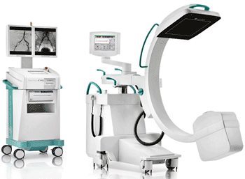
Image: The Ziehm Vision RFD mobile C-arm (Photo courtesy of Ziehm Imaging).
A novel mobile C-arm offers a cost-efficient alternative to fixed installed systems without reducing the imaging capabilities.
Ziehm Imaging (Orlando, FL, USA), a developer, manufacturer, and supplier of mobile C-arms, presented its product range at the scientific assembly and annual meeting of the Radiological Society of North America (RSNA) was held November 27-December 2, 2011, in Chicago (IL, USA).
Global Industry Analysts (San Jose, CA, USA), a market report publisher, predicted extensive growth for the fluoroscopy and mobile C-arm market segments that will be substantially driven by the growing number of minimally invasive procedures. By 2017, the market might reach a volume of USD 1.4 billion. “One of our highlights at this year’s RSNA is Ziehm Vision RFD [radiofrequency detector] hybrid edition that opens up hybrid room applications for physicians working in interdisciplinary environments,” said Nelson Mendes, president and CEO Ziehm Imaging, Inc.
The hybrid edition features the innovative flat-panel technology and delivers high-resolution images with over 16,000 shades of gray and a resolution of 1,500 x 1,500 pixels. The detector is not affected by magnetic fields making it ideal for interventional radiologic procedures; it delivers distortion-free, high-resolution images even when close to magnetic resonance (MR) scanners. The squared display format creates a considerably larger visible area with up to 60% more information per X-ray than conventional image intensifiers.
“The image quality is comparable to many fixed imaging systems. Very often, I forget that I am using a mobile system,” stated Grayson H. Wheatley III, MD, from the Arizona Heart Institute (Phoenix, USA). “I perform percutaneous or catheter-based interventions, on complex aortic problems, such as aortic aneurysms. One of the more innovative procedures I perform…combine the advantages of traditional open surgery with image-guided, catheter-based interventions.”
The Ziehm Vision RFD is precisely customized to hybrid operating room (OR) use criteria. With up to 25 images per second, its powerful monoblock generator also produces high-quality X-rays of moving objects such as beating hearts. The C-arm features a user-friendly touchscreen interface that is mounted to the sterile OR table, an injector interface that synchronizes contrast media with the imaging process, and an interface for external displays. The large C-arm opening enables easier positioning and patient access during the intervention.
This next-generation C-arm also comes with the new software SmartVascular, which enables surgeons to create a digital subtraction angiography (DSA) at any time without having to use the touchscreen. It automates all process steps from DSA to road mapping, allowing vascular procedures to be planned with minimum amounts of contrast media and shorter fluoroscopy times.
The powerful 20 kW monoblock generator with a liquid cooling system that enables almost unlimited fluoroscopy time is now also available for the Ziehm Vision R. This innovative technology allows for completely new imaging possibilities for mobile C-arms with image intensifier in vascular surgery. With its rotating anode, the generator uses a variable pulse width of 4 and 50 ms to produce crystal-clear images. Up to 25 frames per second enable high-quality X-rays of vascular structures with optimum contrast.
The Advanced Active Cooling system keeps the operating temperature steady, prevents the system from overheating, and ensures uninterrupted imaging. Dr. Wheatley confirmed, “the C-arm is often used for long and complex cases in the OR which can take hours to complete. The Ziehm Imaging system holds up quite well and has never shut down due to inadequate heat management. This is critical since the last thing we want to worry about when treating a patient is the C-arm shutting down because of overheating.”
Related Links:
Ziehm Imaging
Latest Radiography News
- RSNA AI Challenge Models Can Independently Interpret Mammograms
- New Technique Combines X-Ray Imaging and Radar for Safer Cancer Diagnosis
- New AI Tool Helps Doctors Read Chest X‑Rays Better
- Wearable X-Ray Imaging Detecting Fabric to Provide On-The-Go Diagnostic Scanning
- AI Helps Radiologists Spot More Lesions in Mammograms
- AI Detects Fatty Liver Disease from Chest X-Rays
- AI Detects Hidden Heart Disease in Existing CT Chest Scans
- Ultra-Lightweight AI Model Runs Without GPU to Break Barriers in Lung Cancer Diagnosis
- AI Radiology Tool Identifies Life-Threatening Conditions in Milliseconds

- Machine Learning Algorithm Identifies Cardiovascular Risk from Routine Bone Density Scans
- AI Improves Early Detection of Interval Breast Cancers
- World's Largest Class Single Crystal Diamond Radiation Detector Opens New Possibilities for Diagnostic Imaging
- AI-Powered Imaging Technique Shows Promise in Evaluating Patients for PCI
- Higher Chest X-Ray Usage Catches Lung Cancer Earlier and Improves Survival
- AI-Powered Mammograms Predict Cardiovascular Risk
- Generative AI Model Significantly Reduces Chest X-Ray Reading Time
Channels
MRI
view channel
AI-Assisted Model Enhances MRI Heart Scans
A cardiac MRI can reveal critical information about the heart’s function and any abnormalities, but traditional scans take 30 to 90 minutes and often suffer from poor image quality due to patient movement.... Read more
AI Model Outperforms Doctors at Identifying Patients Most At-Risk of Cardiac Arrest
Hypertrophic cardiomyopathy is one of the most common inherited heart conditions and a leading cause of sudden cardiac death in young individuals and athletes. While many patients live normal lives, some... Read moreUltrasound
view channel
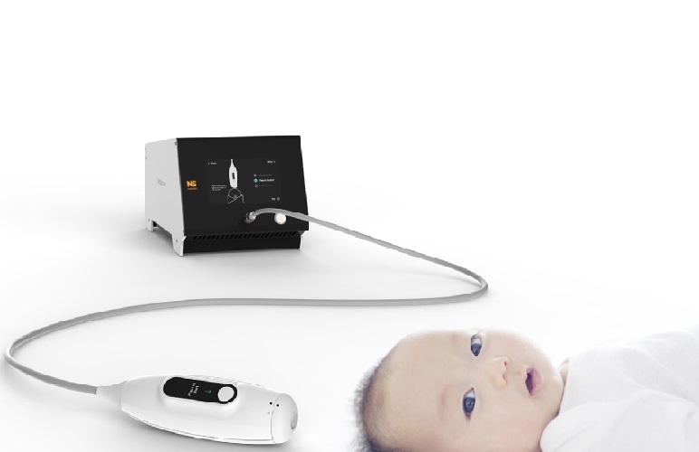
Non-Invasive Ultrasound-Based Tool Accurately Detects Infant Meningitis
Meningitis, an inflammation of the membranes surrounding the brain and spinal cord, can be fatal in infants if not diagnosed and treated early. Even when treated, it may leave lasting damage, such as cognitive... Read more
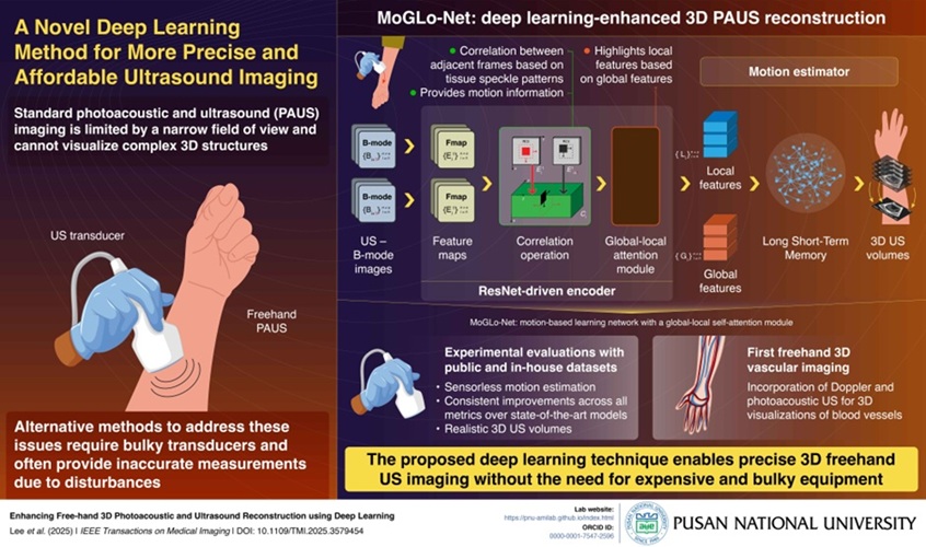
Breakthrough Deep Learning Model Enhances Handheld 3D Medical Imaging
Ultrasound imaging is a vital diagnostic technique used to visualize internal organs and tissues in real time and to guide procedures such as biopsies and injections. When paired with photoacoustic imaging... Read moreNuclear Medicine
view channel
Novel Bacteria-Specific PET Imaging Approach Detects Hard-To-Diagnose Lung Infections
Mycobacteroides abscessus is a rapidly growing mycobacteria that primarily affects immunocompromised patients and those with underlying lung diseases, such as cystic fibrosis or chronic obstructive pulmonary... Read more
New Imaging Approach Could Reduce Need for Biopsies to Monitor Prostate Cancer
Prostate cancer is the second leading cause of cancer-related death among men in the United States. However, the majority of older men diagnosed with prostate cancer have slow-growing, low-risk forms of... Read moreGeneral/Advanced Imaging
view channel
CT Colonography Beats Stool DNA Testing for Colon Cancer Screening
As colorectal cancer remains the second leading cause of cancer-related deaths worldwide, early detection through screening is vital to reduce advanced-stage treatments and associated costs.... Read more
First-Of-Its-Kind Wearable Device Offers Revolutionary Alternative to CT Scans
Currently, patients with conditions such as heart failure, pneumonia, or respiratory distress often require multiple imaging procedures that are intermittent, disruptive, and involve high levels of radiation.... Read more
AI-Based CT Scan Analysis Predicts Early-Stage Kidney Damage Due to Cancer Treatments
Radioligand therapy, a form of targeted nuclear medicine, has recently gained attention for its potential in treating specific types of tumors. However, one of the potential side effects of this therapy... Read moreImaging IT
view channel
New Google Cloud Medical Imaging Suite Makes Imaging Healthcare Data More Accessible
Medical imaging is a critical tool used to diagnose patients, and there are billions of medical images scanned globally each year. Imaging data accounts for about 90% of all healthcare data1 and, until... Read more
Global AI in Medical Diagnostics Market to Be Driven by Demand for Image Recognition in Radiology
The global artificial intelligence (AI) in medical diagnostics market is expanding with early disease detection being one of its key applications and image recognition becoming a compelling consumer proposition... Read moreIndustry News
view channel
GE HealthCare and NVIDIA Collaboration to Reimagine Diagnostic Imaging
GE HealthCare (Chicago, IL, USA) has entered into a collaboration with NVIDIA (Santa Clara, CA, USA), expanding the existing relationship between the two companies to focus on pioneering innovation in... Read more
Patient-Specific 3D-Printed Phantoms Transform CT Imaging
New research has highlighted how anatomically precise, patient-specific 3D-printed phantoms are proving to be scalable, cost-effective, and efficient tools in the development of new CT scan algorithms... Read more
Siemens and Sectra Collaborate on Enhancing Radiology Workflows
Siemens Healthineers (Forchheim, Germany) and Sectra (Linköping, Sweden) have entered into a collaboration aimed at enhancing radiologists' diagnostic capabilities and, in turn, improving patient care... Read more


 Guided Devices.jpg)


