Magnetic Particle Image Technology Provides Real-Time Images of Blood Flow and the Heart
|
By MedImaging International staff writers Posted on 19 Mar 2009 |

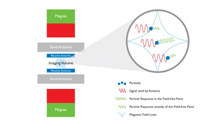
Image: Schematic set up and operating principle of the Magnetic Particle Imaging (MPI) technology (Photo courtesy of Royal Philips Electronics).
New technology called magnetic particle imaging (MPI) has been shown to generate unprecedented real-time images of blood flow and heart movement, which may improve disease diagnosis and treatment planning.
Philips Healthcare (Best, The Netherlands) reported the first three-dimensional (3D) imaging results obtained with MPI. The technology, which uses the magnetic properties of iron-oxide nanoparticles injected into the bloodstream, has been used in a preclinical study to generate extraordinary real-time images of arterial blood flow and volumetric heart motion. This represents a major step forward in taking MPI from a hypothetical concept to an imaging tool to help improve diagnosis and therapy planning for many of the world's major diseases, such as heart disease, stroke, and cancer. The results of the preclinical study were published in March 7, 2009, issue of the journal Physics in Medicine and Biology.
"A novel noninvasive cardiac imaging technology is required to further unravel and characterize the disease processes associated with atherosclerosis, in particular those associated with vulnerable plaque formation, which is a major risk factor for stroke and heart attacks,” said Prof. Valentin Fuster, M.D., Ph.D., director of the Mount Sinai Heart Center (New York, NY, USA). "Through its combined speed, resolution, and sensitivity, magnetic particle imaging technology has great potential for this application, and the latest in vivo imaging results represent a major breakthrough.”
"We are the first in the world to demonstrate that magnetic particle imaging can be used to produce real-time in vivo images that accurately capture cardiovascular activity,” stated Henk van Houten, senior vice president of Philips Research and head of the Healthcare research program. "By adding important functional information to the anatomical data obtained from existing modalities such as CT [computed tomography] and MR [magnetic resonance], Philips' MPI technology has the potential to significantly help in the diagnosis and treatment planning of major diseases such as atherosclerosis and congenital heart defects.”
Philips' magnetic particle imaging utilizes the magnetic properties of injected iron-oxide nanoparticles to measure the nanoparticle concentration in the blood. Because the human body contains no naturally occurring magnetic materials visible to MPI, there is no background signal. After injection, the nanoparticles therefore appear as bright signals in the images, from which nanoparticle concentrations can be calculated. By combining high spatial resolution with short image acquisition times (as short as 1/50th of a second), MPI can capture dynamic concentration changes as the nanoparticles are swept along by the blood stream. This could ultimately allow MPI scanners to perform a wide range of functional cardiovascular measurements in a single scan. These could include measurements of coronary blood supply, myocardial perfusion, and the heart's ejection fraction, wall motion, and flow speeds.
The results obtained from Philips' experimental MPI scanner marks an important step towards the development of a whole-body system for use on humans. Some of the technical challenges in scaling up the system relate to the magnetic field generation required for human applications. Others lie in the measurement and processing of the extremely weak signal emitted by the nanoparticles.
Related Links:
Philips Healthcare
Latest Radiography News
- Machine Learning Algorithm Identifies Cardiovascular Risk from Routine Bone Density Scans
- AI Improves Early Detection of Interval Breast Cancers
- World's Largest Class Single Crystal Diamond Radiation Detector Opens New Possibilities for Diagnostic Imaging
- AI-Powered Imaging Technique Shows Promise in Evaluating Patients for PCI
- Higher Chest X-Ray Usage Catches Lung Cancer Earlier and Improves Survival
- AI-Powered Mammograms Predict Cardiovascular Risk
- Generative AI Model Significantly Reduces Chest X-Ray Reading Time
- AI-Powered Mammography Screening Boosts Cancer Detection in Single-Reader Settings
- Photon Counting Detectors Promise Fast Color X-Ray Images
- AI Can Flag Mammograms for Supplemental MRI
- 3D CT Imaging from Single X-Ray Projection Reduces Radiation Exposure
- AI Method Accurately Predicts Breast Cancer Risk by Analyzing Multiple Mammograms
- Printable Organic X-Ray Sensors Could Transform Treatment for Cancer Patients
- Highly Sensitive, Foldable Detector to Make X-Rays Safer
- Novel Breast Cancer Screening Technology Could Offer Superior Alternative to Mammogram
- Artificial Intelligence Accurately Predicts Breast Cancer Years Before Diagnosis
Channels
MRI
view channel
Simple Brain Scan Diagnoses Parkinson's Disease Years Before It Becomes Untreatable
Parkinson's disease (PD) remains a challenging condition to treat, with no known cure. Though therapies have improved over time, and ongoing research focuses on methods to slow or alter the disease’s progression,... Read more
Cutting-Edge MRI Technology to Revolutionize Diagnosis of Common Heart Problem
Aortic stenosis is a common and potentially life-threatening heart condition. It occurs when the aortic valve, which regulates blood flow from the heart to the rest of the body, becomes stiff and narrow.... Read moreUltrasound
view channel
New Incision-Free Technique Halts Growth of Debilitating Brain Lesions
Cerebral cavernous malformations (CCMs), also known as cavernomas, are abnormal clusters of blood vessels that can grow in the brain, spinal cord, or other parts of the body. While most cases remain asymptomatic,... Read more.jpeg)
AI-Powered Lung Ultrasound Outperforms Human Experts in Tuberculosis Diagnosis
Despite global declines in tuberculosis (TB) rates in previous years, the incidence of TB rose by 4.6% from 2020 to 2023. Early screening and rapid diagnosis are essential elements of the World Health... Read moreNuclear Medicine
view channel
Novel Radiolabeled Antibody Improves Diagnosis and Treatment of Solid Tumors
Interleukin-13 receptor α-2 (IL13Rα2) is a cell surface receptor commonly found in solid tumors such as glioblastoma, melanoma, and breast cancer. It is minimally expressed in normal tissues, making it... Read more
Novel PET Imaging Approach Offers Never-Before-Seen View of Neuroinflammation
COX-2, an enzyme that plays a key role in brain inflammation, can be significantly upregulated by inflammatory stimuli and neuroexcitation. Researchers suggest that COX-2 density in the brain could serve... Read moreGeneral/Advanced Imaging
view channel
AI-Based CT Scan Analysis Predicts Early-Stage Kidney Damage Due to Cancer Treatments
Radioligand therapy, a form of targeted nuclear medicine, has recently gained attention for its potential in treating specific types of tumors. However, one of the potential side effects of this therapy... Read more
CT-Based Deep Learning-Driven Tool to Enhance Liver Cancer Diagnosis
Medical imaging, such as computed tomography (CT) scans, plays a crucial role in oncology, offering essential data for cancer detection, treatment planning, and monitoring of response to therapies.... Read moreImaging IT
view channel
New Google Cloud Medical Imaging Suite Makes Imaging Healthcare Data More Accessible
Medical imaging is a critical tool used to diagnose patients, and there are billions of medical images scanned globally each year. Imaging data accounts for about 90% of all healthcare data1 and, until... Read more
Global AI in Medical Diagnostics Market to Be Driven by Demand for Image Recognition in Radiology
The global artificial intelligence (AI) in medical diagnostics market is expanding with early disease detection being one of its key applications and image recognition becoming a compelling consumer proposition... Read moreIndustry News
view channel
GE HealthCare and NVIDIA Collaboration to Reimagine Diagnostic Imaging
GE HealthCare (Chicago, IL, USA) has entered into a collaboration with NVIDIA (Santa Clara, CA, USA), expanding the existing relationship between the two companies to focus on pioneering innovation in... Read more
Patient-Specific 3D-Printed Phantoms Transform CT Imaging
New research has highlighted how anatomically precise, patient-specific 3D-printed phantoms are proving to be scalable, cost-effective, and efficient tools in the development of new CT scan algorithms... Read more
Siemens and Sectra Collaborate on Enhancing Radiology Workflows
Siemens Healthineers (Forchheim, Germany) and Sectra (Linköping, Sweden) have entered into a collaboration aimed at enhancing radiologists' diagnostic capabilities and, in turn, improving patient care... Read more


