Findings of Zika Virus Effects Detailed in Special Imaging Report
|
By MedImaging International staff writers Posted on 29 Aug 2016 |

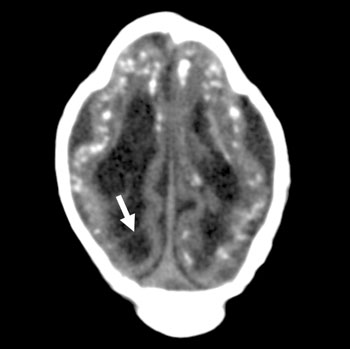
Image: The image displays an axial CT scan of a 1-month-old male with a presumed Zika virus infection, and shows ventriculomegaly with septation, calcifications, an abnormally diffused gyral pattern, and a deformed skull (Photo courtesy of RSNA).
Researchers in Brazil have released imaging findings of the effects of the Zika virus on babies and fetuses infected with the virus.
The researchers used Computed Tomography (CT), Magnetic Resonance Imaging (MRI), and ultrasound imaging to investigate the wide array of effects on the brain, including microcephaly.
The report was published in the August 2016 issue of the journal Radiology. The researchers used imaging and autopsy findings from babies and fetuses affected by the congenital Zika virus infection, and treated at the Northeast Brazilian Instituto de Pesquisa in Campina Grande state Paraiba (IPESQ). The researchers performed a retrospective review of the CT, MRI, and ultrasound images collected between June 2015 and May 2016. The researchers used images and related data of 17 fetuses or neonates of women who had been scanned at IPESQ, and 28 other fetuses or neonates with brain findings that were suspicious for Zika infections.
The researchers found various brain abnormalities in fetuses exposed to the Zika virus, besides microcephaly. These included loss of volume in the gray matter and white matter of the brain, abnormalities of the brainstem, ventriculomegaly, and calcifications.
Lead author of the report, Fernanda Tovar-Moll, MD, PhD, said, "Imaging is essential for identifying the presence and the severity of the structural changes induced by the infection, especially in the central nervous system. Microcephaly is just one of several radiological features. The severity of the cortical malformation and associated tissue changes, and the localization of the calcifications at the grey-white matter junction were the most surprising findings in our research. More than one ultrasound or MRI scan in pregnancy may be needed to assess the growth and development abnormalities of the brain. We are also interested in investigating how congenital Zika virus infection can interfere with not only prenatal, but also postnatal gray and white brain maturation."
Related Links:
RSNA
Latest Ultrasound News
- Diagnostic System Automatically Analyzes TTE Images to Identify Congenital Heart Disease
- Super-Resolution Imaging Technique Could Improve Evaluation of Cardiac Conditions

- First AI-Powered POC Ultrasound Diagnostic Solution Helps Prioritize Cases Based On Severity

- Largest Model Trained On Echocardiography Images Assesses Heart Structure and Function
- Groundbreaking Technology Enables Precise, Automatic Measurement of Peripheral Blood Vessels
- Deep Learning Advances Super-Resolution Ultrasound Imaging
- Novel Ultrasound-Launched Targeted Nanoparticle Eliminates Biofilm and Bacterial Infection
- AI-Guided Ultrasound System Enables Rapid Assessments of DVT
- Focused Ultrasound Technique Gets Quality Assurance Protocol
- AI-Guided Handheld Ultrasound System Helps Capture Diagnostic-Quality Cardiac Images
- Non-Invasive Ultrasound Imaging Device Diagnoses Risk of Chronic Kidney Disease
- Wearable Ultrasound Platform Paves Way for 24/7 Blood Pressure Monitoring On the Wrist
- Diagnostic Ultrasound Enhancing Agent to Improve Image Quality in Pediatric Heart Patients
- AI Detects COVID-19 in Lung Ultrasound Images
- New Ultrasound Technology to Revolutionize Respiratory Disease Diagnoses
- Dynamic Contrast-Enhanced Ultrasound Highly Useful For Interventions
Channels
MRI
view channel
Low-Cost Whole-Body MRI Device Combined with AI Generates High-Quality Results
Magnetic Resonance Imaging (MRI) has significantly transformed healthcare, providing a noninvasive, radiation-free method for detailed imaging. It is especially promising for the future of medical diagnosis... Read more
World's First Whole-Body Ultra-High Field MRI Officially Comes To Market
The world's first whole-body ultra-high field (UHF) MRI has officially come to market, marking a remarkable advancement in diagnostic radiology. United Imaging (Shanghai, China) has secured clearance from the U.... Read moreUltrasound
view channel.jpg)
Diagnostic System Automatically Analyzes TTE Images to Identify Congenital Heart Disease
Congenital heart disease (CHD) is one of the most prevalent congenital anomalies worldwide, presenting substantial health and financial challenges for affected patients. Early detection and treatment of... Read more
Super-Resolution Imaging Technique Could Improve Evaluation of Cardiac Conditions
The heart depends on efficient blood circulation to pump blood throughout the body, delivering oxygen to tissues and removing carbon dioxide and waste. Yet, when heart vessels are damaged, it can disrupt... Read more
First AI-Powered POC Ultrasound Diagnostic Solution Helps Prioritize Cases Based On Severity
Ultrasound scans are essential for identifying and diagnosing various medical conditions, but often, patients must wait weeks or months for results due to a shortage of qualified medical professionals... Read moreNuclear Medicine
view channelNew PET Agent Rapidly and Accurately Visualizes Lesions in Clear Cell Renal Cell Carcinoma Patients
Clear cell renal cell cancer (ccRCC) represents 70-80% of renal cell carcinoma cases. While localized disease can be effectively treated with surgery and ablative therapies, one-third of patients either... Read more
New Imaging Technique Monitors Inflammation Disorders without Radiation Exposure
Imaging inflammation using traditional radiological techniques presents significant challenges, including radiation exposure, poor image quality, high costs, and invasive procedures. Now, new contrast... Read more
New SPECT/CT Technique Could Change Imaging Practices and Increase Patient Access
The development of lead-212 (212Pb)-PSMA–based targeted alpha therapy (TAT) is garnering significant interest in treating patients with metastatic castration-resistant prostate cancer. The imaging of 212Pb,... Read moreGeneral/Advanced Imaging
view channelBone Density Test Uses Existing CT Images to Predict Fractures
Osteoporotic fractures are not only devastating and deadly, especially hip fractures, but also impose significant costs. They rank among the top chronic diseases in terms of disability-adjusted life years... Read more
AI Predicts Cardiac Risk and Mortality from Routine Chest CT Scans
Heart disease remains the leading cause of death and is largely preventable, yet many individuals are unaware of their risk until it becomes severe. Early detection through screening can reveal heart issues,... Read moreImaging IT
view channel
New Google Cloud Medical Imaging Suite Makes Imaging Healthcare Data More Accessible
Medical imaging is a critical tool used to diagnose patients, and there are billions of medical images scanned globally each year. Imaging data accounts for about 90% of all healthcare data1 and, until... Read more
Global AI in Medical Diagnostics Market to Be Driven by Demand for Image Recognition in Radiology
The global artificial intelligence (AI) in medical diagnostics market is expanding with early disease detection being one of its key applications and image recognition becoming a compelling consumer proposition... Read moreIndustry News
view channel
Hologic Acquires UK-Based Breast Surgical Guidance Company Endomagnetics Ltd.
Hologic, Inc. (Marlborough, MA, USA) has entered into a definitive agreement to acquire Endomagnetics Ltd. (Cambridge, UK), a privately held developer of breast cancer surgery technologies, for approximately... Read more
Bayer and Google Partner on New AI Product for Radiologists
Medical imaging data comprises around 90% of all healthcare data, and it is a highly complex and rich clinical data modality and serves as a vital tool for diagnosing patients. Each year, billions of medical... Read more


