Novel Fluorescent Imaging Agent Helps Surgeons Visualize Missed Lung Tumors
|
By MedImaging International staff writers Posted on 24 Jul 2024 |

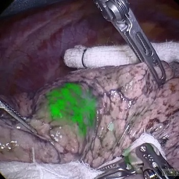
Surgery remains the primary treatment for most solid tumors, but conventional open surgery often results in significant trauma and high costs. Minimally invasive (MIS) and robotic-assisted surgical techniques have become increasingly popular as they reduce tissue damage, pain, blood loss, duration of hospital stays, and postoperative complications. Despite these advantages, these advanced surgical methods can sometimes leave behind tumor tissue, which is linked to poorer patient outcomes, including reduced survival rates. Now, a new study has shown that a novel tumor-targeted fluorescent imaging agent enables surgeons to better see and remove tumor tissues that are typically missed using standard surgical approaches.
Vergent Bioscience’s (Minneapolis, MN, USA) VGT-309 is an innovative tumor-targeted fluorescent imaging agent intended to enhance tumor visibility during open, MIS, and robotic-assisted surgeries. Administered through a brief intravenous infusion a few hours before surgery, VGT-309 tightly binds to cathepsins—proteases overexpressed in many solid tumors. This binding property potentially offers significant clinical benefits, making VGT-309 an optimal imaging agent for tumor detection. VGT-309 incorporates the near infrared (NIR) dye indocyanine green (ICG), which is compatible with all current NIR intraoperative imaging systems designed for MIS. ICG is favored for its ability to minimize background autofluorescence that can interfere with imaging clarity.
Now, data published in The Annals of Thoracic Surgery further validate the clinical utility of VGT-309, demonstrating its capability to reveal difficult-to-find and previously undetected tumors in lung surgeries. In a Phase 2 efficacy study involving 40 patients with suspected or confirmed lung cancer eligible for surgery, researchers assessed whether IMI with VGT-309 could enhance surgical outcomes. The study's primary efficacy endpoint was the rate of patients experiencing at least one clinically significant event, defined as the detection of lesions overlooked by standard techniques, the discovery of synchronous and occult cancers, and the identification of inadequate surgical margins.
Patients received VGT-309 preoperatively via intravenous infusion. After attempting to locate the lesion using standard techniques, surgeons employed a commercially available NIR endoscope to visualize the lesion, which was then evaluated by pathology. Out of the 40 participants treated with VGT-309, 17 (42.5%) experienced at least one clinically significant event during their surgery. The use of VGT-309 with NIR fluorescence imaging enabled intraoperative visualization of various primary and metastatic tumors, including adenocarcinoma in situ, invasive adenocarcinoma, lymphoma, colorectal cancer, neuroendocrine tumors, sarcomas, and squamous cell carcinoma. VGT-309 proved safe and well-tolerated in this study, with no infusion reactions or drug-related serious adverse events reported. These results support previous clinical findings, suggesting that VGT-309 could play a crucial role in ensuring the complete removal of tumor tissue during MIS and robotic-assisted surgeries.
“The expanded use of minimally invasive surgery, including robotic-assisted technologies, for lung cancer has created a significant and growing need for better visualization during these surgeries, which can be curative when lung cancer is diagnosed early enough and all tumor tissue is removed,” said John Santini, Ph.D., president and chief executive officer at Vergent Bioscience. “We are pleased that data from our clinical program continue to suggest VGT-309 may help overcome existing challenges to tumor visualization and thereby optimize surgical outcomes for physicians and patients.”
Related Links:
Vergent Bioscience
Latest Nuclear Medicine News
- Novel Radiolabeled Antibody Improves Diagnosis and Treatment of Solid Tumors
- Novel PET Imaging Approach Offers Never-Before-Seen View of Neuroinflammation
- Novel Radiotracer Identifies Biomarker for Triple-Negative Breast Cancer
- Innovative PET Imaging Technique to Help Diagnose Neurodegeneration
- New Molecular Imaging Test to Improve Lung Cancer Diagnosis
- Novel PET Technique Visualizes Spinal Cord Injuries to Predict Recovery
- Next-Gen Tau Radiotracers Outperform FDA-Approved Imaging Agents in Detecting Alzheimer’s
- Breakthrough Method Detects Inflammation in Body Using PET Imaging
- Advanced Imaging Reveals Hidden Metastases in High-Risk Prostate Cancer Patients
- Combining Advanced Imaging Technologies Offers Breakthrough in Glioblastoma Treatment
- New Molecular Imaging Agent Accurately Identifies Crucial Cancer Biomarker
- New Scans Light Up Aggressive Tumors for Better Treatment
- AI Stroke Brain Scan Readings Twice as Accurate as Current Method
- AI Analysis of PET/CT Images Predicts Side Effects of Immunotherapy in Lung Cancer
- New Imaging Agent to Drive Step-Change for Brain Cancer Imaging
- Portable PET Scanner to Detect Earliest Stages of Alzheimer’s Disease
Channels
Radiography
view channel
World's Largest Class Single Crystal Diamond Radiation Detector Opens New Possibilities for Diagnostic Imaging
Diamonds possess ideal physical properties for radiation detection, such as exceptional thermal and chemical stability along with a quick response time. Made of carbon with an atomic number of six, diamonds... Read more
AI-Powered Imaging Technique Shows Promise in Evaluating Patients for PCI
Percutaneous coronary intervention (PCI), also known as coronary angioplasty, is a minimally invasive procedure where small metal tubes called stents are inserted into partially blocked coronary arteries... Read moreMRI
view channel
AI Tool Predicts Relapse of Pediatric Brain Cancer from Brain MRI Scans
Many pediatric gliomas are treatable with surgery alone, but relapses can be catastrophic. Predicting which patients are at risk for recurrence remains challenging, leading to frequent follow-ups with... Read more
AI Tool Tracks Effectiveness of Multiple Sclerosis Treatments Using Brain MRI Scans
Multiple sclerosis (MS) is a condition in which the immune system attacks the brain and spinal cord, leading to impairments in movement, sensation, and cognition. Magnetic Resonance Imaging (MRI) markers... Read more
Ultra-Powerful MRI Scans Enable Life-Changing Surgery in Treatment-Resistant Epileptic Patients
Approximately 360,000 individuals in the UK suffer from focal epilepsy, a condition in which seizures spread from one part of the brain. Around a third of these patients experience persistent seizures... Read moreUltrasound
view channel.jpeg)
AI-Powered Lung Ultrasound Outperforms Human Experts in Tuberculosis Diagnosis
Despite global declines in tuberculosis (TB) rates in previous years, the incidence of TB rose by 4.6% from 2020 to 2023. Early screening and rapid diagnosis are essential elements of the World Health... Read more
AI Identifies Heart Valve Disease from Common Imaging Test
Tricuspid regurgitation is a condition where the heart's tricuspid valve does not close completely during contraction, leading to backward blood flow, which can result in heart failure. A new artificial... Read moreNuclear Medicine
view channel
Novel Radiolabeled Antibody Improves Diagnosis and Treatment of Solid Tumors
Interleukin-13 receptor α-2 (IL13Rα2) is a cell surface receptor commonly found in solid tumors such as glioblastoma, melanoma, and breast cancer. It is minimally expressed in normal tissues, making it... Read more
Novel PET Imaging Approach Offers Never-Before-Seen View of Neuroinflammation
COX-2, an enzyme that plays a key role in brain inflammation, can be significantly upregulated by inflammatory stimuli and neuroexcitation. Researchers suggest that COX-2 density in the brain could serve... Read moreGeneral/Advanced Imaging
view channel
AI-Powered Imaging System Improves Lung Cancer Diagnosis
Given the need to detect lung cancer at earlier stages, there is an increasing need for a definitive diagnostic pathway for patients with suspicious pulmonary nodules. However, obtaining tissue samples... Read more
AI Model Significantly Enhances Low-Dose CT Capabilities
Lung cancer remains one of the most challenging diseases, making early diagnosis vital for effective treatment. Fortunately, advancements in artificial intelligence (AI) are revolutionizing lung cancer... Read moreImaging IT
view channel
New Google Cloud Medical Imaging Suite Makes Imaging Healthcare Data More Accessible
Medical imaging is a critical tool used to diagnose patients, and there are billions of medical images scanned globally each year. Imaging data accounts for about 90% of all healthcare data1 and, until... Read more
Global AI in Medical Diagnostics Market to Be Driven by Demand for Image Recognition in Radiology
The global artificial intelligence (AI) in medical diagnostics market is expanding with early disease detection being one of its key applications and image recognition becoming a compelling consumer proposition... Read moreIndustry News
view channel
GE HealthCare and NVIDIA Collaboration to Reimagine Diagnostic Imaging
GE HealthCare (Chicago, IL, USA) has entered into a collaboration with NVIDIA (Santa Clara, CA, USA), expanding the existing relationship between the two companies to focus on pioneering innovation in... Read more
Patient-Specific 3D-Printed Phantoms Transform CT Imaging
New research has highlighted how anatomically precise, patient-specific 3D-printed phantoms are proving to be scalable, cost-effective, and efficient tools in the development of new CT scan algorithms... Read more
Siemens and Sectra Collaborate on Enhancing Radiology Workflows
Siemens Healthineers (Forchheim, Germany) and Sectra (Linköping, Sweden) have entered into a collaboration aimed at enhancing radiologists' diagnostic capabilities and, in turn, improving patient care... Read more


