New Use for Copper in MRI Contrast Agent Design Enables Clearer Images and Improved Diagnosis
|
By MedImaging International staff writers Posted on 12 Jul 2023 |

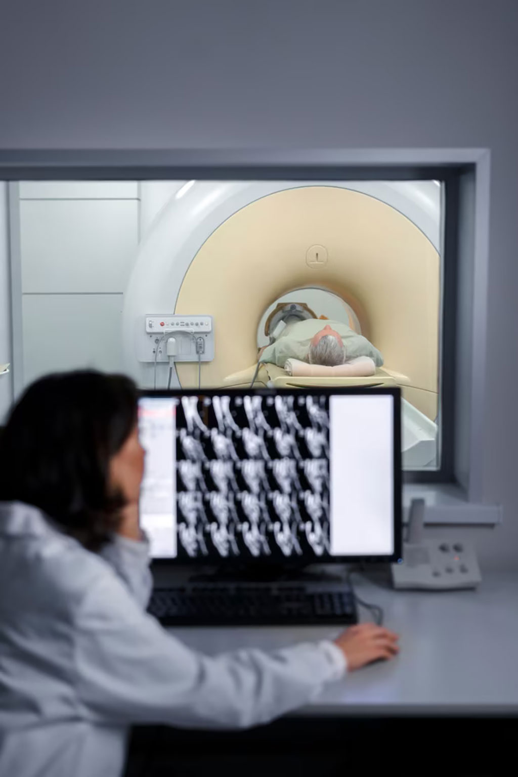
Magnetic resonance imaging (MRI) scanners illuminate portions of the body using a strong magnetic field, leading to hydrogen nuclei of water in tissues being polarized in the direction of the magnetic field. The magnitude of the spin polarization is measured to generate MR images but decays according to a specific time constant known as the T1 relaxation time. Water protons in different tissues exhibit varied T1 values, serving as a primary source of contrast in MR images. The T1 value of nearby water protons can be both reduced and, occasionally, increased through the use of a contrast agent, enhancing the image contrast and thereby boosting the clarity of internal bodily structures. The most common compounds used for this purpose are gadolinium-based contrast agents (GBCAs). While Gadolinium (in the form of Gd3+) is frequently employed as a contrast agent, environmental and patient safety concerns have led to the ongoing search for new contrast agents.
In a groundbreaking research collaboration, scientists at the University of Birmingham (Birmingham, UK) have uncovered a novel application of copper in designing MRI contrast agents. This finding holds promise for generating superior, safer images that facilitate easier and safer patient diagnosis. The researchers identified a novel copper protein binding site that holds significant potential for use in MRI contrast agents to enhance the visibility of internal body structures. This discovery defies the traditional belief that copper is ill-suited for MRI contrast agents and may contribute to the creation of new imaging agents posing fewer risks and side effects than those currently in use.
The research team succeeded in creating a highly elusive abiological copper site bound to oxygen donor atoms within a protein scaffold. They found that this new structure exhibited high levels of relaxivity - the capacity of a contrast agent to affect the proton relaxation times, leading to clearer and more detailed MRI images. The researchers suggest that copper-based imaging agents may also be used in Positron emission tomography (PET) scans, which generate intricate 3D internal body images. Their study highlights how the creation of a copper site within a protein scaffold using an artificial coiled coil resulted in functionality and performance not typically linked to copper.
“We prepared a new-to-biology copper–binding site which shows real potential for use in contrast agents and challenges existing dogma that copper is unsuitable for use in MRI,” said co-author Dr Anna Peacock, Reader in Bioinorganic Chemistry at the University of Birmingham. “Despite copper largely being disregarded for use in MRI contrast agents, our binding site was shown to display extremely promising contrast agent capabilities, with relaxivities equal and superior to the Gd(III) agents used routinely in clinical MRI. Our discovery showcases a powerful approach for accessing new tools or agents for imaging applications.”
Related Links:
University of Birmingham
Latest General/Advanced Imaging News
- CT-Based Deep Learning-Driven Tool to Enhance Liver Cancer Diagnosis
- AI-Powered Imaging System Improves Lung Cancer Diagnosis
- AI Model Significantly Enhances Low-Dose CT Capabilities
- Ultra-Low Dose CT Aids Pneumonia Diagnosis in Immunocompromised Patients
- AI Reduces CT Lung Cancer Screening Workload by Almost 80%
- Cutting-Edge Technology Combines Light and Sound for Real-Time Stroke Monitoring
- AI System Detects Subtle Changes in Series of Medical Images Over Time
- New CT Scan Technique to Improve Prognosis and Treatments for Head and Neck Cancers
- World’s First Mobile Whole-Body CT Scanner to Provide Diagnostics at POC
- Comprehensive CT Scans Could Identify Atherosclerosis Among Lung Cancer Patients
- AI Improves Detection of Colorectal Cancer on Routine Abdominopelvic CT Scans
- Super-Resolution Technology Enhances Clinical Bone Imaging to Predict Osteoporotic Fracture Risk
- AI-Powered Abdomen Map Enables Early Cancer Detection
- Deep Learning Model Detects Lung Tumors on CT
- AI Predicts Cardiovascular Risk from CT Scans
- Deep Learning Based Algorithms Improve Tumor Detection in PET/CT Scans
Channels
Radiography
view channel
AI Improves Early Detection of Interval Breast Cancers
Interval breast cancers, which occur between routine screenings, are easier to treat when detected earlier. Early detection can reduce the need for aggressive treatments and improve the chances of better outcomes.... Read more
World's Largest Class Single Crystal Diamond Radiation Detector Opens New Possibilities for Diagnostic Imaging
Diamonds possess ideal physical properties for radiation detection, such as exceptional thermal and chemical stability along with a quick response time. Made of carbon with an atomic number of six, diamonds... Read moreUltrasound
view channel.jpeg)
AI-Powered Lung Ultrasound Outperforms Human Experts in Tuberculosis Diagnosis
Despite global declines in tuberculosis (TB) rates in previous years, the incidence of TB rose by 4.6% from 2020 to 2023. Early screening and rapid diagnosis are essential elements of the World Health... Read more
AI Identifies Heart Valve Disease from Common Imaging Test
Tricuspid regurgitation is a condition where the heart's tricuspid valve does not close completely during contraction, leading to backward blood flow, which can result in heart failure. A new artificial... Read moreNuclear Medicine
view channel
Novel Radiolabeled Antibody Improves Diagnosis and Treatment of Solid Tumors
Interleukin-13 receptor α-2 (IL13Rα2) is a cell surface receptor commonly found in solid tumors such as glioblastoma, melanoma, and breast cancer. It is minimally expressed in normal tissues, making it... Read more
Novel PET Imaging Approach Offers Never-Before-Seen View of Neuroinflammation
COX-2, an enzyme that plays a key role in brain inflammation, can be significantly upregulated by inflammatory stimuli and neuroexcitation. Researchers suggest that COX-2 density in the brain could serve... Read moreGeneral/Advanced Imaging
view channel
CT-Based Deep Learning-Driven Tool to Enhance Liver Cancer Diagnosis
Medical imaging, such as computed tomography (CT) scans, plays a crucial role in oncology, offering essential data for cancer detection, treatment planning, and monitoring of response to therapies.... Read more
AI-Powered Imaging System Improves Lung Cancer Diagnosis
Given the need to detect lung cancer at earlier stages, there is an increasing need for a definitive diagnostic pathway for patients with suspicious pulmonary nodules. However, obtaining tissue samples... Read moreImaging IT
view channel
New Google Cloud Medical Imaging Suite Makes Imaging Healthcare Data More Accessible
Medical imaging is a critical tool used to diagnose patients, and there are billions of medical images scanned globally each year. Imaging data accounts for about 90% of all healthcare data1 and, until... Read more
Global AI in Medical Diagnostics Market to Be Driven by Demand for Image Recognition in Radiology
The global artificial intelligence (AI) in medical diagnostics market is expanding with early disease detection being one of its key applications and image recognition becoming a compelling consumer proposition... Read moreIndustry News
view channel
GE HealthCare and NVIDIA Collaboration to Reimagine Diagnostic Imaging
GE HealthCare (Chicago, IL, USA) has entered into a collaboration with NVIDIA (Santa Clara, CA, USA), expanding the existing relationship between the two companies to focus on pioneering innovation in... Read more
Patient-Specific 3D-Printed Phantoms Transform CT Imaging
New research has highlighted how anatomically precise, patient-specific 3D-printed phantoms are proving to be scalable, cost-effective, and efficient tools in the development of new CT scan algorithms... Read more
Siemens and Sectra Collaborate on Enhancing Radiology Workflows
Siemens Healthineers (Forchheim, Germany) and Sectra (Linköping, Sweden) have entered into a collaboration aimed at enhancing radiologists' diagnostic capabilities and, in turn, improving patient care... Read more


