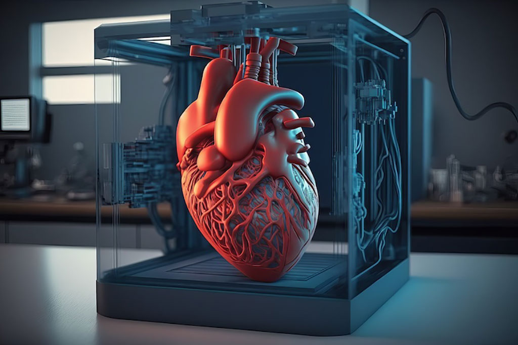
3D Printed Heart Models Could Enable Non-Invasive Diagnosis of Aortic Stenosis
|
By MedImaging International staff writers Posted on 11 Apr 2023 |

Aortic stenosis is a condition characterized by calcified and thickened aortic heart valves, which impede blood flow. Existing methods for assessing the severity of aortic stenosis, like Doppler echocardiography, can be prone to uncontrolled errors and often necessitate invasive pressure measurements for patients. No, aortic flow phantoms could provide a potential solution to this issue.
Researchers at King’s College London (KCL, London, UK) have made progress in utilizing 3D printed heart models (phantoms) to simulate and investigate aortic stenosis. Computer modeling and 3D printing of aortic flow phantoms present an alternative to in vivo studies, which are associated with challenges in patient recruitment and potential procedural risks. In contrast, the simulated option allows for greater variations in blood pressure flow and drop. The researchers created a non-invasive technique for evaluating pressure recovery distance based on blood flow momentum using 4D Flow cardiovascular magnetic resonance (CMR). Their findings revealed that pressure recovery distances in aortic stenosis are longer than previously recognized, indicating a need to reevaluate currently adopted interventional practices.
Furthermore, the researchers developed and successfully tested a flow phantom compatible with MRI and ultrasound, which accurately simulates valve opening and closing in both healthy and diseased conditions and offers ground-truth pressure measurement. The team's findings suggest that the peak-to-peak pressure drop, a current metric for assessing the burden of aortic stenosis, may be influenced by factors unrelated to the valve, such as wave reflection, and should be reexamined in clinical practice.
“By developing valve models that behave like real human valves, new techniques which more accurately characterize the severity of disease can be developed and improved without disrupting patients' care,” said Harminder Gill, BM BCh.
“The decision on how and when to treat stenotic valves is complex and the diagnostic tools typically used in clinical routine have barely evolved during the past 50 years,” explained Joao Filipe Fernandes, PhD, Marie Skłodowska-Curie Early Stage Researcher in Personalized in-Silico Cardiology. “Thus, advances in the study of aortic stenosis patho-physiology are essential to provide a more comprehensive characterization of this condition. The non-invasive assessment of the pressure recovery distance allows the detection of invasive catheterization errors as well as understanding the vessel length required for hemodynamic homeostasis to be reached.”
“These advances will enable us to take well informed decision on the best balance between drugs and surgeries for people living with valve conditions,” added Prof. Pablo Lamata, Head of Cardiac Modeling and Imaging Biomarkers Group.
Related Links:
King’s College London
Latest MRI News
- New Material Boosts MRI Image Quality
- AI Model Reads and Diagnoses Brain MRI in Seconds
- MRI Scan Breakthrough to Help Avoid Risky Invasive Tests for Heart Patients
- MRI Scans Reveal Signature Patterns of Brain Activity to Predict Recovery from TBI
- Novel Imaging Approach to Improve Treatment for Spinal Cord Injuries
- AI-Assisted Model Enhances MRI Heart Scans
- AI Model Outperforms Doctors at Identifying Patients Most At-Risk of Cardiac Arrest
- New MRI Technique Reveals Hidden Heart Issues
- Shorter MRI Exam Effectively Detects Cancer in Dense Breasts
- MRI to Replace Painful Spinal Tap for Faster MS Diagnosis
- MRI Scans Can Identify Cardiovascular Disease Ten Years in Advance
- Simple Brain Scan Diagnoses Parkinson's Disease Years Before It Becomes Untreatable
- Cutting-Edge MRI Technology to Revolutionize Diagnosis of Common Heart Problem
- New MRI Technique Reveals True Heart Age to Prevent Attacks and Strokes
- AI Tool Predicts Relapse of Pediatric Brain Cancer from Brain MRI Scans
- AI Tool Tracks Effectiveness of Multiple Sclerosis Treatments Using Brain MRI Scans
Channels
Radiography
view channel
Routine Mammograms Could Predict Future Cardiovascular Disease in Women
Mammograms are widely used to screen for breast cancer, but they may also contain overlooked clues about cardiovascular health. Calcium deposits in the arteries of the breast signal stiffening blood vessels,... Read more
AI Detects Early Signs of Aging from Chest X-Rays
Chronological age does not always reflect how fast the body is truly aging, and current biological age tests often rely on DNA-based markers that may miss early organ-level decline. Detecting subtle, age-related... Read moreUltrasound
view channel
Reusable Gel Pad Made from Tamarind Seed Could Transform Ultrasound Examinations
Ultrasound imaging depends on a conductive gel to eliminate air between the probe and the skin so sound waves can pass clearly into the body. While the imaging technology is fast, safe, and noninvasive,... Read more
AI Model Accurately Detects Placenta Accreta in Pregnancy Before Delivery
Placenta accreta spectrum (PAS) is a life-threatening pregnancy complication in which the placenta abnormally attaches to the uterine wall. The condition is a leading cause of maternal mortality and morbidity... Read moreNuclear Medicine
view channel
Radiopharmaceutical Molecule Marker to Improve Choice of Bladder Cancer Therapies
Targeted cancer therapies only work when tumor cells express the specific molecular structures they are designed to attack. In urothelial carcinoma, a common form of bladder cancer, the cell surface protein... Read more
Cancer “Flashlight” Shows Who Can Benefit from Targeted Treatments
Targeted cancer therapies can be highly effective, but only when a patient’s tumor expresses the specific protein the treatment is designed to attack. Determining this usually requires biopsies or advanced... Read moreGeneral/Advanced Imaging
view channel
AI Tool Offers Prognosis for Patients with Head and Neck Cancer
Oropharyngeal cancer is a form of head and neck cancer that can spread through lymph nodes, significantly affecting survival and treatment decisions. Current therapies often involve combinations of surgery,... Read more
New 3D Imaging System Addresses MRI, CT and Ultrasound Limitations
Medical imaging is central to diagnosing and managing injuries, cancer, infections, and chronic diseases, yet existing tools each come with trade-offs. Ultrasound, X-ray, CT, and MRI can be costly, time-consuming,... Read moreImaging IT
view channel
New Google Cloud Medical Imaging Suite Makes Imaging Healthcare Data More Accessible
Medical imaging is a critical tool used to diagnose patients, and there are billions of medical images scanned globally each year. Imaging data accounts for about 90% of all healthcare data1 and, until... Read more
Global AI in Medical Diagnostics Market to Be Driven by Demand for Image Recognition in Radiology
The global artificial intelligence (AI) in medical diagnostics market is expanding with early disease detection being one of its key applications and image recognition becoming a compelling consumer proposition... Read moreIndustry News
view channel
Nuclear Medicine Set for Continued Growth Driven by Demand for Precision Diagnostics
Clinical imaging services face rising demand for precise molecular diagnostics and targeted radiopharmaceutical therapy as cancer and chronic disease rates climb. A new market analysis projects rapid expansion... Read more


 Guided Devices.jpg)


