Ultrasound-Guided Surgery More Effective for Treating Early Form of Breast Cancer
|
By MedImaging International staff writers Posted on 21 Nov 2022 |

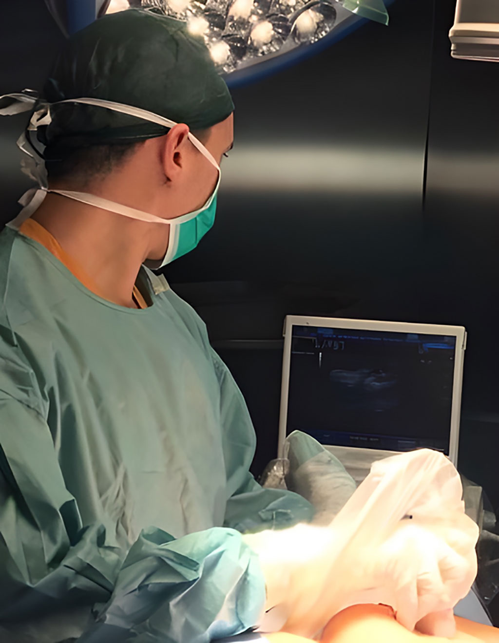
Using ultrasound to guide surgery for patients with ductal carcinoma in situ (DCIS) gives better results than the standard technique of using a wire inserted into the breast, according to new research. The technique, known as intraoperative ultrasound, or IOUS, enables surgeons to remove a smaller quantity of breast tissue, while still removing all the of DCIS tissue. Using IOUS improves the chance of having no cancer cells at the outer edge of the tissue that was removed (known as positive margins), reducing the risk of patients needing a second operation. Because IOUS negates the need for inserting a guide wire, this technique could also reduce pain and preoperative stress for patients and save time for medical staff.
The research carried out by a team at Clinica Universidad de Navarra (Madrid, Spain) involved 108 people who were diagnosed with DCIS and treated between February 2018 and December 2021. Forty-one were treated with IOUS-guided surgery while 67 were treated with surgery guided by wire localization (WL). Following each operation, the tissue removed was analyzed to see how much was removed and whether there were ‘positive margins’. This means that DCIS cells were found at the edge of the tissue removed, suggesting some DCIS cells could have been left behind and patients would probably need a second operation.
Among those treated using WL, seven (10.4%) had positive margins and needed a second operation while in those treated using IOUS, there were only two patients (4.8%) with positive margins who needed a second operation. The patients have been followed up for a year and half so far and cancer has only recurred in one (who was treated with WL). The researchers plan to continue gathering information about patients having surgery for DCIS in the hopes of seeing long-term benefits of using IOUS.
“DCIS is a common form of early breast cancer that can develop into a more serious, invasive cancer. To ensure it does not progress, patients are usually offered surgery. Because DCIS does not usually create lumps in the breast, we need a good technique to guide surgery and make it accurate as possible,” said Dr. Antonio J. Esgueva at Clinica Universidad de Navarra. “As breast surgeons, we want to perform the very best oncological surgery in terms of removing any trace of DCIS but also removing as little of the breast tissue as possible in order to have the best cosmetic result possible. At the same time, we also want to improve patients' experience during treatment by using less invasive techniques and reducing their anxiety. Our research suggests that using intraoperative ultrasound, a quicker and less invasive technique, is effective for guiding DCIS surgery.”
“Once intervention is planned, the standard treatment for patients diagnosed with DCIS is surgery. The need for a second operation due to positive margins can be an issue. This research is promising because it shows that a kinder technique can help guide surgeons to effectively remove DCIS from the breast while minimising unwanted side-effects,” added Dr. Laura Biganzoli, Co-Chair of the European Breast Cancer Conference and Director of the Breast Centre at Santo Stefano Hospital, Prato, Italy, who was not involved in the research.
Related Links:
Clinica Universidad de Navarra
Latest Ultrasound News
- AI-Powered Lung Ultrasound Outperforms Human Experts in Tuberculosis Diagnosis
- AI Identifies Heart Valve Disease from Common Imaging Test
- Novel Imaging Method Enables Early Diagnosis and Treatment Monitoring of Type 2 Diabetes
- Ultrasound-Based Microscopy Technique to Help Diagnose Small Vessel Diseases
- Smart Ultrasound-Activated Immune Cells Destroy Cancer Cells for Extended Periods
- Tiny Magnetic Robot Takes 3D Scans from Deep Within Body
- High Resolution Ultrasound Speeds Up Prostate Cancer Diagnosis
- World's First Wireless, Handheld, Whole-Body Ultrasound with Single PZT Transducer Makes Imaging More Accessible
- Artificial Intelligence Detects Undiagnosed Liver Disease from Echocardiograms
- Ultrasound Imaging Non-Invasively Tracks Tumor Response to Radiation and Immunotherapy
- AI Improves Detection of Congenital Heart Defects on Routine Prenatal Ultrasounds
- AI Diagnoses Lung Diseases from Ultrasound Videos with 96.57% Accuracy
- New Contrast Agent for Ultrasound Imaging Ensures Affordable and Safer Medical Diagnostics
- Ultrasound-Directed Microbubbles Boost Immune Response Against Tumors
- POC Ultrasound Enhances Early Pregnancy Care and Cuts Emergency Visits
- AI-Based Models Outperform Human Experts at Identifying Ovarian Cancer in Ultrasound Images
Channels
Radiography
view channel
World's Largest Class Single Crystal Diamond Radiation Detector Opens New Possibilities for Diagnostic Imaging
Diamonds possess ideal physical properties for radiation detection, such as exceptional thermal and chemical stability along with a quick response time. Made of carbon with an atomic number of six, diamonds... Read more
AI-Powered Imaging Technique Shows Promise in Evaluating Patients for PCI
Percutaneous coronary intervention (PCI), also known as coronary angioplasty, is a minimally invasive procedure where small metal tubes called stents are inserted into partially blocked coronary arteries... Read moreMRI
view channel
AI Tool Predicts Relapse of Pediatric Brain Cancer from Brain MRI Scans
Many pediatric gliomas are treatable with surgery alone, but relapses can be catastrophic. Predicting which patients are at risk for recurrence remains challenging, leading to frequent follow-ups with... Read more
AI Tool Tracks Effectiveness of Multiple Sclerosis Treatments Using Brain MRI Scans
Multiple sclerosis (MS) is a condition in which the immune system attacks the brain and spinal cord, leading to impairments in movement, sensation, and cognition. Magnetic Resonance Imaging (MRI) markers... Read more
Ultra-Powerful MRI Scans Enable Life-Changing Surgery in Treatment-Resistant Epileptic Patients
Approximately 360,000 individuals in the UK suffer from focal epilepsy, a condition in which seizures spread from one part of the brain. Around a third of these patients experience persistent seizures... Read moreNuclear Medicine
view channel
Novel Radiolabeled Antibody Improves Diagnosis and Treatment of Solid Tumors
Interleukin-13 receptor α-2 (IL13Rα2) is a cell surface receptor commonly found in solid tumors such as glioblastoma, melanoma, and breast cancer. It is minimally expressed in normal tissues, making it... Read more
Novel PET Imaging Approach Offers Never-Before-Seen View of Neuroinflammation
COX-2, an enzyme that plays a key role in brain inflammation, can be significantly upregulated by inflammatory stimuli and neuroexcitation. Researchers suggest that COX-2 density in the brain could serve... Read moreGeneral/Advanced Imaging
view channel
AI-Powered Imaging System Improves Lung Cancer Diagnosis
Given the need to detect lung cancer at earlier stages, there is an increasing need for a definitive diagnostic pathway for patients with suspicious pulmonary nodules. However, obtaining tissue samples... Read more
AI Model Significantly Enhances Low-Dose CT Capabilities
Lung cancer remains one of the most challenging diseases, making early diagnosis vital for effective treatment. Fortunately, advancements in artificial intelligence (AI) are revolutionizing lung cancer... Read moreImaging IT
view channel
New Google Cloud Medical Imaging Suite Makes Imaging Healthcare Data More Accessible
Medical imaging is a critical tool used to diagnose patients, and there are billions of medical images scanned globally each year. Imaging data accounts for about 90% of all healthcare data1 and, until... Read more
Global AI in Medical Diagnostics Market to Be Driven by Demand for Image Recognition in Radiology
The global artificial intelligence (AI) in medical diagnostics market is expanding with early disease detection being one of its key applications and image recognition becoming a compelling consumer proposition... Read moreIndustry News
view channel
GE HealthCare and NVIDIA Collaboration to Reimagine Diagnostic Imaging
GE HealthCare (Chicago, IL, USA) has entered into a collaboration with NVIDIA (Santa Clara, CA, USA), expanding the existing relationship between the two companies to focus on pioneering innovation in... Read more
Patient-Specific 3D-Printed Phantoms Transform CT Imaging
New research has highlighted how anatomically precise, patient-specific 3D-printed phantoms are proving to be scalable, cost-effective, and efficient tools in the development of new CT scan algorithms... Read more
Siemens and Sectra Collaborate on Enhancing Radiology Workflows
Siemens Healthineers (Forchheim, Germany) and Sectra (Linköping, Sweden) have entered into a collaboration aimed at enhancing radiologists' diagnostic capabilities and, in turn, improving patient care... Read more


 Guided Devices.jpg)


