New Contrast Agent for Ultrasound Imaging Ensures Affordable and Safer Medical Diagnostics
Posted on 22 Jan 2025
Ultrasound imaging is an affordable and non-invasive diagnostic method that uses widely available equipment. However, its results are often not highly accurate, and the image quality is heavily dependent on the operator's skill. In contrast, techniques like MRI and CT scans are not always accessible due to their high cost, lack of available equipment, and potential harm caused by the contrast agents used in these methods. These contrast agents are toxic, and CT scans expose patients to high radiation doses. On the other hand, ultrasound imaging with contrast agents can significantly improve the quality of images for internal organs, such as the heart, liver, and brain. This enhanced ultrasound technique could be useful for diagnosing a wide array of diseases in the cardiovascular, liver, kidney, and female reproductive systems. Ultrasound contrast agents are often microbubbles made from proteins, lipids, or polymers. Proteins enhance image quality, while lipids and polymers provide stability, extending the time available for diagnostics. Despite these benefits, further improvements in their performance are still needed.
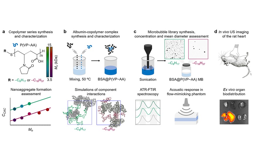
Researchers at Skolkovo Institute of Science and Technology (Skoltech, Moscow, Russia) have developed and tested protein-polymer microbubbles for medical ultrasound imaging. When injected intravenously, these microbubbles serve as contrast agents, improving image quality. Tested in rats, the new microbubbles stayed in the bloodstream longer than the currently available alternatives, giving doctors more time to complete the examination. This enhanced contrast could make ultrasound a viable alternative to MRI and CT scans, which are more expensive and potentially harmful. The researchers created the first "hybrid" microbubble formulation by combining a protein called albumin with a copolymer.

This formulation resulted in 100 different microbubble types for ultrasound imaging, from which the most effective contrast agent was chosen and tested on a rat’s beating heart. The selection process involved multiple steps: evaluating microbubble size, concentration, and acoustic response in organ phantoms and animals. Compared to a protein-based agent without the added copolymer, the hybrid microbubbles showed significant improvements in both image contrast and the time they remained in the bloodstream, which was one and a half to two times longer.
To create the new contrast agent, the team combined bovine serum albumin—a commonly used protein in pharmacology, including in microbubble production—with biocompatible polymers. They tested 100 different combinations, varying the type of polymer and the polymer's proportion in the mixture from 2% to nearly 50%. The challenge was to find the right balance between microbubble stability, concentration, and ultrasound response. Formulations that did not produce stable microbubbles were discarded, and the remaining candidates were tested on a blood vessel phantom to measure bubble size, concentration, and acoustic response. This process led to the identification of the most promising formulation, which was then compared with pure albumin microbubbles and a saline injection in animal trials.
In these tests, the contrast agent was injected into a rat’s tail vein, followed by an ultrasound exam of its heart. The results, published in Biomaterials Advances, revealed that while the image without contrast showed few details, and the image with the protein contrast only displayed the ventricles, the hybrid agent provided a clear view of the entire four-chamber heart. Moreover, the effect lasted up to 7 minutes, compared to just 3 minutes with standard agents. The researchers believe the new agent’s high contrast could make it a valuable tool for diagnosing infertility in women and studying blood vessels in the brain, although further work is needed to fully explore its potential.


