Novel Method Combines Optical Measurements with Ultrasound for Monitoring ‘Engine’ of Pregnancy
|
By MedImaging International staff writers Posted on 17 Aug 2022 |

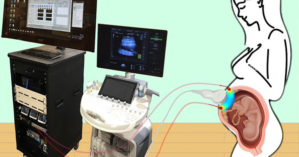
The placenta, considered as the “engine” of pregnancy, is an organ that plays a crucial role in delivering nutrients and oxygen to the fetus. Placental dysfunction can lead to complications such as fetal growth restriction, preeclampsia, and stillbirth. Now, researchers developing tools to assess human placental structure and function in real time, including optical devices, have come up with a novel method for imaging the placenta in pregnant patients. By combining optical measurements with ultrasound, the team has demonstrated how oxygen levels in the placenta can be monitored noninvasively, providing a new way to generate a better understanding of this complex, crucial organ.
For three years, researchers at the University of Pennsylvania (Philadelphia, PA, USA) and the Children’s Hospital of Philadelphia (CHOP, (Philadelphia, PA, USA) optimized the design of their instrument and tested it in preclinical settings. The process involved integrating optical fibers with ultrasound probes, exploring various ultrasound transducers, and improving the multimodal technology so that measurements were stable, accurate, and reproducible while collecting data at the bedside. The resulting instrumentation now enables researchers to study the anatomy of the placenta while also collecting detailed functional information about placenta blood flow and oxygenation, capabilities that existing commercially devices do not have, the researchers say.
Because the placenta is located far below the body’s surface, one of the key technical challenges addressed by the researchers was reducing background noise in the opto-electronic system. Light is scattered and absorbed when it travels through thick tissues, and the key for success was to reduce background interference so that the small amount of light that penetrates deep into the placenta and then returns is still large enough for a high-quality measurement.
In a pilot clinical study conducted by the researchers, 24 pregnant patients in their third trimester were given supplemental oxygen for a short time period, creating placental hyperoxia. Using the device, the team collected measurements of the placenta’s oxygenated and deoxygenated blood concentrations before and during hyperoxia; the results demonstrated that the device could be used to study placental function in real time. The research also provided new insights into the relationship between blood flow and maternal vascular malperfusion, which occurs when blood flow into the placenta is impeded.
While the device is still in development, the researchers are currently refining their instrument to make it more user-friendly and to allow it to collect data faster. The team is also currently working on larger studies, including recently data from patients during their second trimester, and they are also interested in studying different regions of the placenta. And because there are many unanswered clinical questions about the placenta, the biggest potential of this work for the researchers is in providing a way to start answering those questions.
“What was exciting is that, not only did we get an instrument to probe deeper than commercial devices, but we also obtained an early signal that hyperoxygenation experiments can differentiate a healthy placenta from a diseased placenta,” said Nadav Schwartz from the University of Pennsylvania. “Without being able to study the placenta directly, we are relying on very indirect science. This is a tool that helps us study the underlying physiology of pregnancy so we can more strategically study interventions that can help support good pregnancy outcomes.”
Related Links:
University of Pennsylvania
CHOP
Latest Ultrasound News
- AI-Powered Lung Ultrasound Outperforms Human Experts in Tuberculosis Diagnosis
- AI Identifies Heart Valve Disease from Common Imaging Test
- Novel Imaging Method Enables Early Diagnosis and Treatment Monitoring of Type 2 Diabetes
- Ultrasound-Based Microscopy Technique to Help Diagnose Small Vessel Diseases
- Smart Ultrasound-Activated Immune Cells Destroy Cancer Cells for Extended Periods
- Tiny Magnetic Robot Takes 3D Scans from Deep Within Body
- High Resolution Ultrasound Speeds Up Prostate Cancer Diagnosis
- World's First Wireless, Handheld, Whole-Body Ultrasound with Single PZT Transducer Makes Imaging More Accessible
- Artificial Intelligence Detects Undiagnosed Liver Disease from Echocardiograms
- Ultrasound Imaging Non-Invasively Tracks Tumor Response to Radiation and Immunotherapy
- AI Improves Detection of Congenital Heart Defects on Routine Prenatal Ultrasounds
- AI Diagnoses Lung Diseases from Ultrasound Videos with 96.57% Accuracy
- New Contrast Agent for Ultrasound Imaging Ensures Affordable and Safer Medical Diagnostics
- Ultrasound-Directed Microbubbles Boost Immune Response Against Tumors
- POC Ultrasound Enhances Early Pregnancy Care and Cuts Emergency Visits
- AI-Based Models Outperform Human Experts at Identifying Ovarian Cancer in Ultrasound Images
Channels
Radiography
view channel
AI Improves Early Detection of Interval Breast Cancers
Interval breast cancers, which occur between routine screenings, are easier to treat when detected earlier. Early detection can reduce the need for aggressive treatments and improve the chances of better outcomes.... Read more
World's Largest Class Single Crystal Diamond Radiation Detector Opens New Possibilities for Diagnostic Imaging
Diamonds possess ideal physical properties for radiation detection, such as exceptional thermal and chemical stability along with a quick response time. Made of carbon with an atomic number of six, diamonds... Read moreMRI
view channel
Cutting-Edge MRI Technology to Revolutionize Diagnosis of Common Heart Problem
Aortic stenosis is a common and potentially life-threatening heart condition. It occurs when the aortic valve, which regulates blood flow from the heart to the rest of the body, becomes stiff and narrow.... Read more
New MRI Technique Reveals True Heart Age to Prevent Attacks and Strokes
Heart disease remains one of the leading causes of death worldwide. Individuals with conditions such as diabetes or obesity often experience accelerated aging of their hearts, sometimes by decades.... Read more
AI Tool Predicts Relapse of Pediatric Brain Cancer from Brain MRI Scans
Many pediatric gliomas are treatable with surgery alone, but relapses can be catastrophic. Predicting which patients are at risk for recurrence remains challenging, leading to frequent follow-ups with... Read more
AI Tool Tracks Effectiveness of Multiple Sclerosis Treatments Using Brain MRI Scans
Multiple sclerosis (MS) is a condition in which the immune system attacks the brain and spinal cord, leading to impairments in movement, sensation, and cognition. Magnetic Resonance Imaging (MRI) markers... Read moreNuclear Medicine
view channel
Novel Radiolabeled Antibody Improves Diagnosis and Treatment of Solid Tumors
Interleukin-13 receptor α-2 (IL13Rα2) is a cell surface receptor commonly found in solid tumors such as glioblastoma, melanoma, and breast cancer. It is minimally expressed in normal tissues, making it... Read more
Novel PET Imaging Approach Offers Never-Before-Seen View of Neuroinflammation
COX-2, an enzyme that plays a key role in brain inflammation, can be significantly upregulated by inflammatory stimuli and neuroexcitation. Researchers suggest that COX-2 density in the brain could serve... Read moreGeneral/Advanced Imaging
view channel
CT-Based Deep Learning-Driven Tool to Enhance Liver Cancer Diagnosis
Medical imaging, such as computed tomography (CT) scans, plays a crucial role in oncology, offering essential data for cancer detection, treatment planning, and monitoring of response to therapies.... Read more
AI-Powered Imaging System Improves Lung Cancer Diagnosis
Given the need to detect lung cancer at earlier stages, there is an increasing need for a definitive diagnostic pathway for patients with suspicious pulmonary nodules. However, obtaining tissue samples... Read moreImaging IT
view channel
New Google Cloud Medical Imaging Suite Makes Imaging Healthcare Data More Accessible
Medical imaging is a critical tool used to diagnose patients, and there are billions of medical images scanned globally each year. Imaging data accounts for about 90% of all healthcare data1 and, until... Read more
Global AI in Medical Diagnostics Market to Be Driven by Demand for Image Recognition in Radiology
The global artificial intelligence (AI) in medical diagnostics market is expanding with early disease detection being one of its key applications and image recognition becoming a compelling consumer proposition... Read moreIndustry News
view channel
GE HealthCare and NVIDIA Collaboration to Reimagine Diagnostic Imaging
GE HealthCare (Chicago, IL, USA) has entered into a collaboration with NVIDIA (Santa Clara, CA, USA), expanding the existing relationship between the two companies to focus on pioneering innovation in... Read more
Patient-Specific 3D-Printed Phantoms Transform CT Imaging
New research has highlighted how anatomically precise, patient-specific 3D-printed phantoms are proving to be scalable, cost-effective, and efficient tools in the development of new CT scan algorithms... Read more
Siemens and Sectra Collaborate on Enhancing Radiology Workflows
Siemens Healthineers (Forchheim, Germany) and Sectra (Linköping, Sweden) have entered into a collaboration aimed at enhancing radiologists' diagnostic capabilities and, in turn, improving patient care... Read more


