CT Imaging Alone Found Noninferior to CT/MR Combo for Acute Ischemic Stroke Patients
|
By MedImaging International staff writers Posted on 25 Jul 2022 |


|
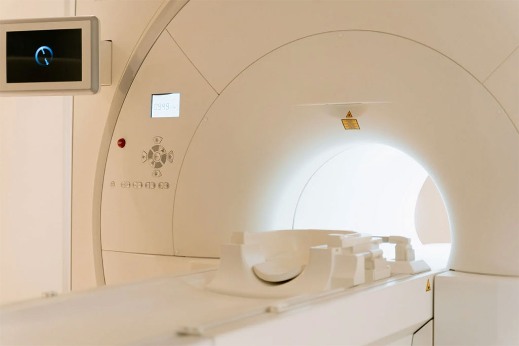
More than 90% of patients with acute ischemic stroke (AIS) receive magnetic resonance imaging (MRI) in addition to computed tomography (CT) with few data to determine whether there is an associated benefit with patient outcomes. Now, a new study of patients hospitalized with AIS has found that a diagnostic imaging strategy of initial CT alone was noninferior to initial CT with additional MRI with regard to the clinical outcomes of death or dependence at hospital discharge or prevention of stroke or death at one year after discharge.
The study by researchers at the Duke University Medical Center (Durham, NC, USA) involved 246 participants with a median age of 68 years and 131 being men (53%). The researchers found that death or dependence at discharge occurred more often in patients with additional MRI (59 of 123 [48%]) than in those with CT alone (52 of 123 [42.3%]); meeting the −7.50% criterion for noninferiority. Stroke or death within one year after discharge determined for 225 of 235 (96%) survivors occurred more often in patients with additional MRI (22 of 113 [19.5%]) than in those with CT alone (14 of 112 [12.5%]), meeting the 0.725 relative risk criterion for noninferiority.
Based on the findings of the propensity score-matched cohort study of patients hospitalized with AIS, the researchers concluded that a diagnostic imaging strategy of initial CT alone was noninferior to initial CT plus additional MRI with regard to clinical outcomes at discharge and at one year. Further research is needed to determine which patients hospitalized with acute ischemic stroke benefit from MRI, according to the researchers.
Related Links:
Duke University Medical Center



 Guided Devices.jpg)


Latest MRI News
- Cardiac MRI Measure Improves Risk Prediction in Tricuspid Regurgitation
- AI System Improves Accuracy of Cardiac MRI Interpretation
- Deep Learning Model Predicts Alzheimer’s Disease Outcomes from Baseline MRI
- Blood-Brain Barrier Imaging Adds Risk Insight to Standard Stroke MRI
- AI Body Composition MRI Analysis Predicts Cardiometabolic Disease Risk
- AI MRI Tool Quantifies Muscle Fat to Assess Cardiometabolic Risk
- Advanced MRI Visualizes CSF Motion Changes After Mild Traumatic Brain Injury
- MRI Tool Enables Long-Term Tracking of Transplanted Cardiac Cells
- MRI-Based AI Tool Supports Differentiation of Parkinsonian Syndromes
- MRI-Derived Biomarker Improves Risk Stratification in Glioblastoma
- Combined Imaging Approach Identifies Cause of Heart Attack without Coronary Blockage
- Advanced MRI System Detects Impaired Cardiac Oxygen Use in Minutes
- AI-Enhanced MRI Improves Image Quality in Arrhythmia Patients
- Ultra-Detailed Brain Atlas Enhances Early Detection of Neurological Disorders
- Study Finds Advanced Imaging Significantly Reduces Unnecessary Prostate Biopsies
- New Material Boosts MRI Image Quality
Channels
Radiography
view channel
Simple Chest X-Ray Measure Predicts Survival After Lung Cancer Surgery
Obstructive ventilatory disorder, marked by airflow limitation that reduces breathing efficiency, increases postoperative risk in patients with lung cancer. Although surgery offers the best chance of cure,... Read more
AI Detection Tool Improves Identification of Lobular Breast Cancer
Breast cancer screening seeks early detection, yet some subtypes remain difficult to visualize on mammography, risking delayed diagnosis. On average, 1 in 20 women worldwide will develop breast cancer,... Read more
MRI
view channel
Cardiac MRI Measure Improves Risk Prediction in Tricuspid Regurgitation
Tricuspid regurgitation, in which blood flows back from the right ventricle into the right atrium, can lead to progressive right-sided heart failure. Clinicians need reliable ways to gauge severity and... Read more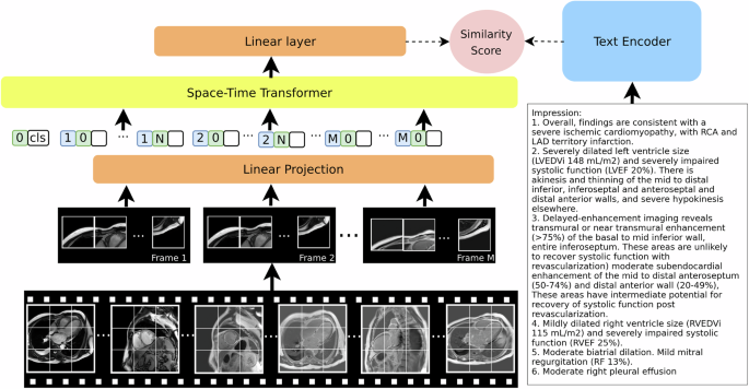
AI System Improves Accuracy of Cardiac MRI Interpretation
Cardiac magnetic resonance imaging (MRI) is the reference standard for assessing cardiac structure, function, and tissue health. Yet each exam can contain hundreds to thousands of images across multiple... Read more
Ultrasound
view channel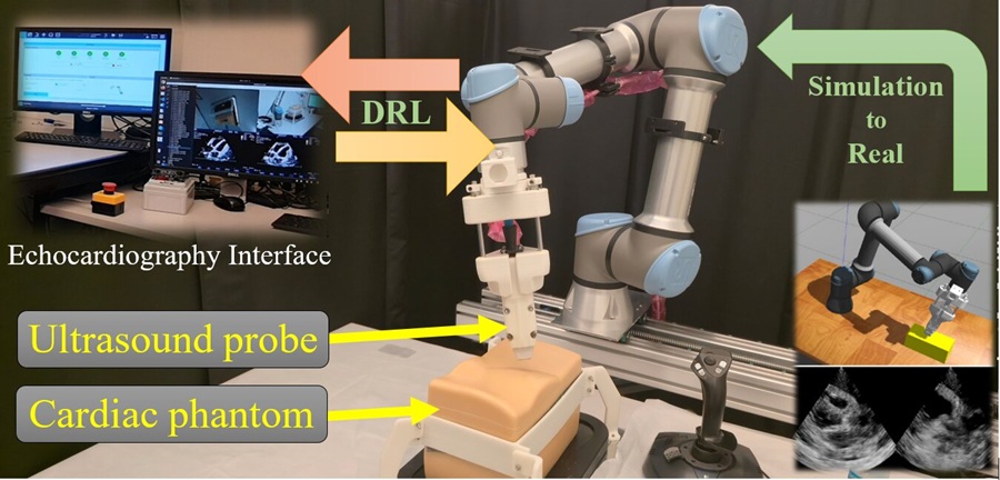
AI Robotic Ultrasound System Automates Echocardiography and Improves Consistency
Echocardiography, an ultrasound examination of the heart, is central to diagnosing and managing cardiovascular disease. Many services struggle with limited availability of skilled sonographers, variable... Read more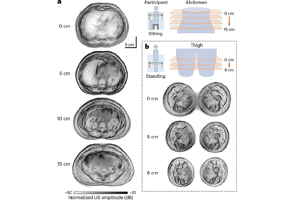
Whole Cross-Section Ultrasound System Enables Operator-Independent Imaging
Conventional ultrasound is central to bedside imaging but is limited by a narrow field of view and operator variability. Comprehensive cross-sectional assessment typically requires computed tomography... Read more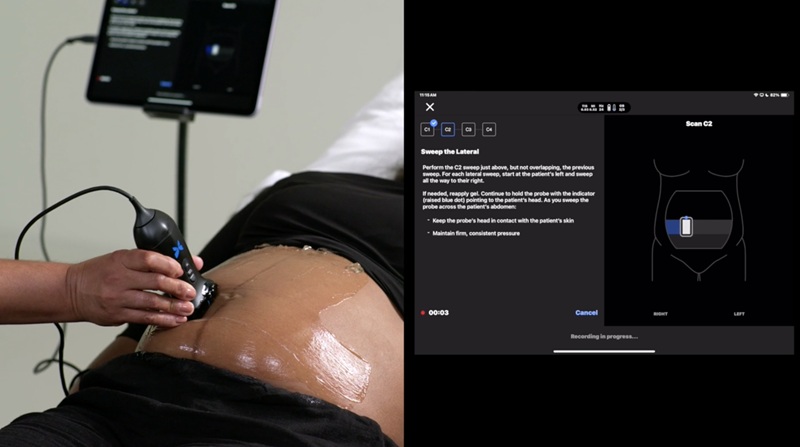
Nuclear Medicine
view channel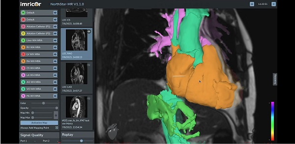
MR-Guided Cardiac Mapping System Enables Radiation-Free Procedures
Cardiac electrophysiology procedures are typically guided by X-ray fluoroscopy, which limits soft-tissue visualization and exposes patients and clinical staff to ionizing radiation. Real-time mapping that... Read more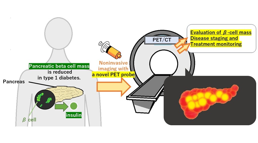
PET Tracer Enables Noninvasive Measurement of Beta Cell Mass
Type 1 diabetes is an autoimmune disease in which the immune system destroys insulin-producing pancreatic beta cells. Loss of these cells destabilizes glucose control and drives complications.... Read more
New Imaging Tool Sheds Light on Tumor Fat Metabolism
Rapidly growing tumors reprogram metabolism to meet high energy demands. While many cancers preferentially consume glucose, lipid utilization by malignant cells is difficult to measure in living subjects.... Read more
Radiopharmaceutical Molecule Marker to Improve Choice of Bladder Cancer Therapies
Targeted cancer therapies only work when tumor cells express the specific molecular structures they are designed to attack. In urothelial carcinoma, a common form of bladder cancer, the cell surface protein... Read moreImaging IT
view channel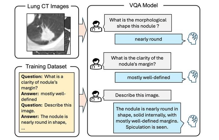
Interactive AI Tool Supports Explainable Lung Nodule Assessment
Lung cancer is a leading cause of cancer mortality, and timely characterization of pulmonary nodules on chest computed tomography (CT) is essential for directing care. Interpreting nodule morphology demands... Read more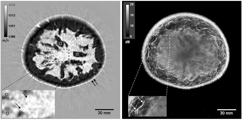
Breast Imaging Software Enhances Visualization and Tissue Characterization in Challenging Cases
Breast imaging can be particularly challenging in cases involving small breasts or implants, where image reconstruction and tissue characterization may be limited. Clinicians also need reproducible analysis... Read more
New Google Cloud Medical Imaging Suite Makes Imaging Healthcare Data More Accessible
Medical imaging is a critical tool used to diagnose patients, and there are billions of medical images scanned globally each year. Imaging data accounts for about 90% of all healthcare data1 and, until... Read more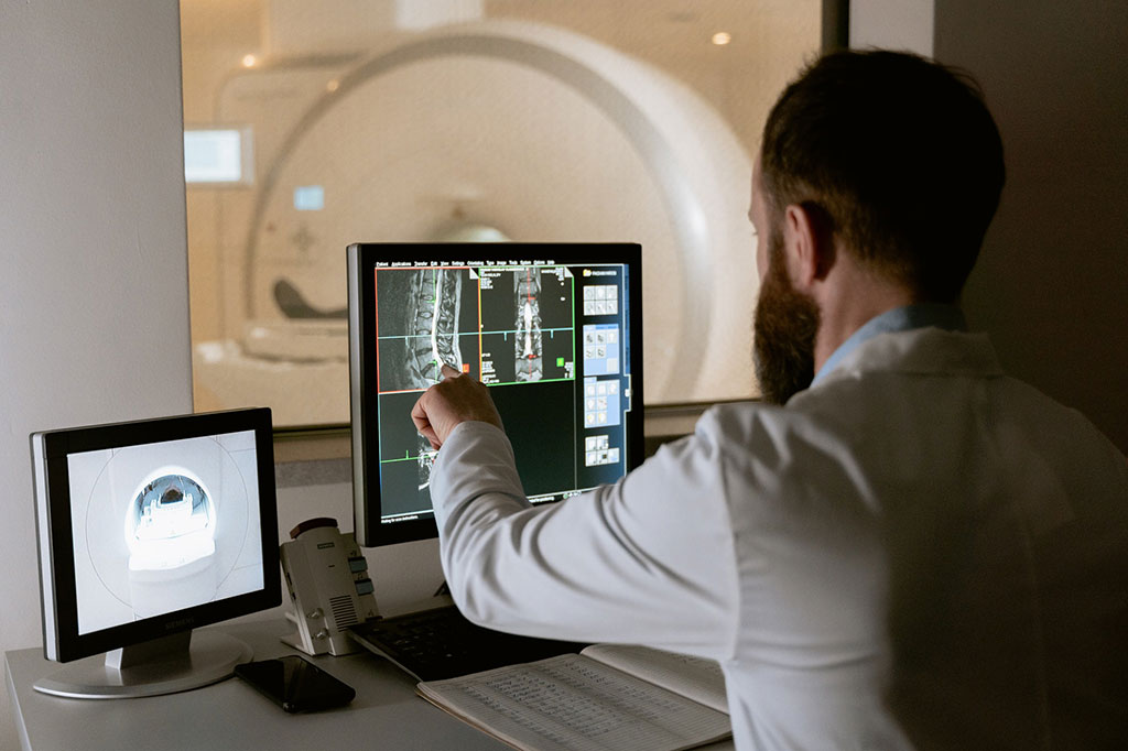
Global AI in Medical Diagnostics Market to Be Driven by Demand for Image Recognition in Radiology
The global artificial intelligence (AI) in medical diagnostics market is expanding with early disease detection being one of its key applications and image recognition becoming a compelling consumer proposition... Read moreIndustry News
view channel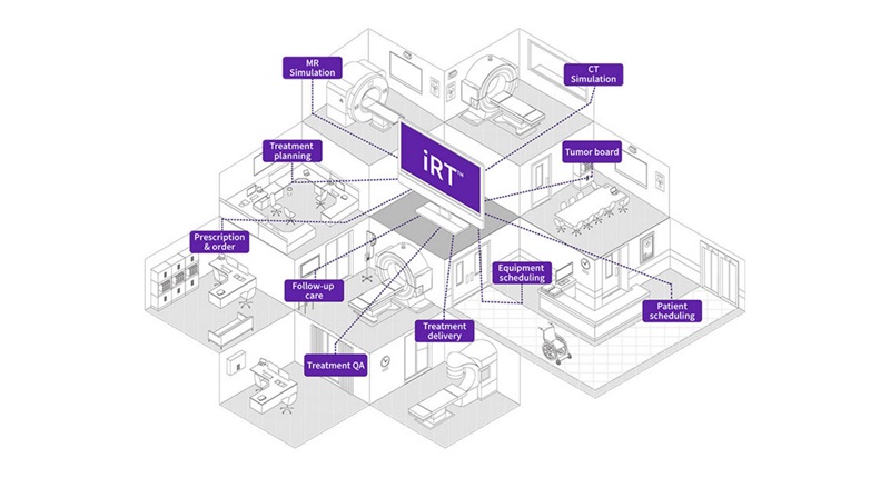
GE HealthCare Highlights AI-Supported Radiation Therapy Tools at ESTRO 2026
At the European Society for Radiotherapy and Oncology (ESTRO) 2026 Congress in Stockholm, GE HealthCare is highlighting Intelligent Radiation Therapy (iRT), MIM Software innovations, and BK Medical surgical... Read more