Artificial Intelligence Enables Early Detection of Arthritis Using HR-pQCT Scans
|
By MedImaging International staff writers Posted on 11 May 2022 |

There are many different types of arthritis, and diagnosing the exact type of inflammatory disease that is affecting a patient’s joints is not always easy. Missing biomarkers currently often make precise classification of the relevant type of arthritis difficult. X-ray images used to aid diagnosis are not completely reliable either, as their two-dimensionality is not precise enough and leaves room for interpretation. This is in addition to the fact that positioning the joint being examined for an X-ray image can be difficult. Now, a team of computer scientists and physicians have succeeded in teaching an artificial neural network to differentiate between rheumatoid arthritis, psoriatic arthritis and healthy joints.
An interdisciplinary research project conducted at Friedrich-Alexander-Universität Erlangen-Nürnberg (FAU, Erlangen, Germany) and Universitätsklinikum Erlangen (Erlangen, Germany) investigated the following questions: Can artificial intelligence (AI) detect various types of arthritis using joint shape patterns? Does this method allow us to make more precise diagnoses in cases of undifferentiated arthritis? Are there certain areas in joints that should be examined in more detail during a diagnosis? To find the answers to its questions, the research team focused its investigations on the metacarpophalangeal joints of the fingers – regions in the body that are very often affected early on in patients with autoimmune diseases such as rheumatoid arthritis or psoriatic arthritis.
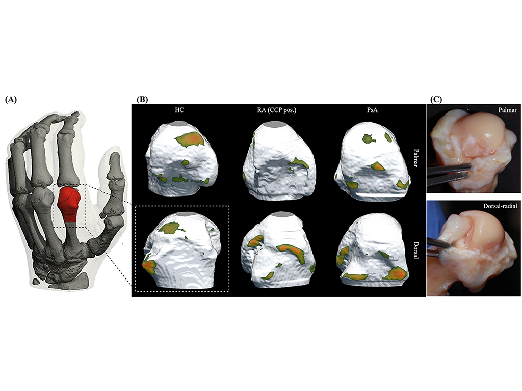
A network of artificial neurons was trained using finger scans from high-resolution peripheral quantitative computer tomography (HR-pQCT) with the aim of differentiating between “healthy” joints and those from patients with rheumatoid or psoriatic arthritis. HR-pQCT was selected as it is currently the best quantitative method of producing three dimensional images of human bones in the highest resolution. In the case of arthritis, changes in the structure of bones can be very accurately detected, which makes precise classification possible. A total of 932 new HR-pQCT scans from 611 patients were then used to check if the artificial network can actually implement what it had learned: Can it provide a correct assessment of the previously classified finger joints?
The results showed that AI detected 82% of the healthy joints, 75% of the cases of rheumatoid arthritis and 68% of the cases of psoriatic arthritis, which is a very high hit probability without any further information. When combined with the expertise of a rheumatologist, it could lead to much more accurate diagnoses. In addition, when presented with cases of undifferentiated arthritis, the network was able to classify them correctly. Whereas the research team was able to use high-resolution computer tomography, this type of imaging is only rarely available to physicians under normal circumstances because of restraints in terms of space and costs. However, these new findings are still useful as the neural network detected certain areas of the joints that provide the most information about a specific type of arthritis that are known as intra-articular hotspots. In the future, physicians could use these areas as another piece in the diagnostic puzzle to confirm suspected cases. This would save time and effort during the diagnosis and is already in fact possible using ultrasound, for example.
“We are very satisfied with the results of the study as they show that artificial intelligence can help us to classify arthritis more easily, which could lead to quicker and more targeted treatment for patients. However, we are aware of the fact that there are other categories that need to be fed into the network. We are also planning to transfer the AI method to other imaging methods such as ultrasound or MRI, which are more readily available,” explained Lukas Folle from the Chair of Computer Science 5 (Pattern Recognition) at Universitätsklinikum Erlangen.
Related Links:
FAU
Universitätsklinikum Erlangen
Latest General/Advanced Imaging News
- AI Tool Offers Prognosis for Patients with Head and Neck Cancer
- New 3D Imaging System Addresses MRI, CT and Ultrasound Limitations
- AI-Based Tool Predicts Future Cardiovascular Events in Angina Patients
- AI-Based Tool Accelerates Detection of Kidney Cancer
- New Algorithm Dramatically Speeds Up Stroke Detection Scans
- 3D Scanning Approach Enables Ultra-Precise Brain Surgery
- AI Tool Improves Medical Imaging Process by 90%
- New Ultrasmall, Light-Sensitive Nanoparticles Could Serve as Contrast Agents
- AI Algorithm Accurately Predicts Pancreatic Cancer Metastasis Using Routine CT Images
- Cutting-Edge Angio-CT Solution Offers New Therapeutic Possibilities
- Extending CT Imaging Detects Hidden Blood Clots in Stroke Patients
- Groundbreaking AI Model Accurately Segments Liver Tumors from CT Scans
- New CT-Based Indicator Helps Predict Life-Threatening Postpartum Bleeding Cases
- CT Colonography Beats Stool DNA Testing for Colon Cancer Screening
- First-Of-Its-Kind Wearable Device Offers Revolutionary Alternative to CT Scans
- AI-Based CT Scan Analysis Predicts Early-Stage Kidney Damage Due to Cancer Treatments
Channels
Radiography
view channel
Routine Mammograms Could Predict Future Cardiovascular Disease in Women
Mammograms are widely used to screen for breast cancer, but they may also contain overlooked clues about cardiovascular health. Calcium deposits in the arteries of the breast signal stiffening blood vessels,... Read more
AI Detects Early Signs of Aging from Chest X-Rays
Chronological age does not always reflect how fast the body is truly aging, and current biological age tests often rely on DNA-based markers that may miss early organ-level decline. Detecting subtle, age-related... Read moreMRI
view channel
New Material Boosts MRI Image Quality
Magnetic resonance imaging (MRI) is a cornerstone of modern diagnostics, yet certain deep or anatomically complex tissues, including delicate structures of the eye and orbit, remain difficult to visualize clearly.... Read more
AI Model Reads and Diagnoses Brain MRI in Seconds
Brain MRI scans are critical for diagnosing strokes, hemorrhages, and other neurological disorders, but interpreting them can take hours or even days due to growing demand and limited specialist availability.... Read moreMRI Scan Breakthrough to Help Avoid Risky Invasive Tests for Heart Patients
Heart failure patients often require right heart catheterization to assess how severely their heart is struggling to pump blood, a procedure that involves inserting a tube into the heart to measure blood... Read more
MRI Scans Reveal Signature Patterns of Brain Activity to Predict Recovery from TBI
Recovery after traumatic brain injury (TBI) varies widely, with some patients regaining full function while others are left with lasting disabilities. Prognosis is especially difficult to assess in patients... Read moreUltrasound
view channel
Reusable Gel Pad Made from Tamarind Seed Could Transform Ultrasound Examinations
Ultrasound imaging depends on a conductive gel to eliminate air between the probe and the skin so sound waves can pass clearly into the body. While the imaging technology is fast, safe, and noninvasive,... Read more
AI Model Accurately Detects Placenta Accreta in Pregnancy Before Delivery
Placenta accreta spectrum (PAS) is a life-threatening pregnancy complication in which the placenta abnormally attaches to the uterine wall. The condition is a leading cause of maternal mortality and morbidity... Read moreNuclear Medicine
view channel
Radiopharmaceutical Molecule Marker to Improve Choice of Bladder Cancer Therapies
Targeted cancer therapies only work when tumor cells express the specific molecular structures they are designed to attack. In urothelial carcinoma, a common form of bladder cancer, the cell surface protein... Read more
Cancer “Flashlight” Shows Who Can Benefit from Targeted Treatments
Targeted cancer therapies can be highly effective, but only when a patient’s tumor expresses the specific protein the treatment is designed to attack. Determining this usually requires biopsies or advanced... Read moreImaging IT
view channel
New Google Cloud Medical Imaging Suite Makes Imaging Healthcare Data More Accessible
Medical imaging is a critical tool used to diagnose patients, and there are billions of medical images scanned globally each year. Imaging data accounts for about 90% of all healthcare data1 and, until... Read more
Global AI in Medical Diagnostics Market to Be Driven by Demand for Image Recognition in Radiology
The global artificial intelligence (AI) in medical diagnostics market is expanding with early disease detection being one of its key applications and image recognition becoming a compelling consumer proposition... Read moreIndustry News
view channel
Nuclear Medicine Set for Continued Growth Driven by Demand for Precision Diagnostics
Clinical imaging services face rising demand for precise molecular diagnostics and targeted radiopharmaceutical therapy as cancer and chronic disease rates climb. A new market analysis projects rapid expansion... Read more


