AI Predicts Spinal Fractures in Cancer Patients Using CT/MRI Scans
|
By MedImaging International staff writers Posted on 06 May 2022 |

One of the biggest clinical concerns that cancer patients face is the risk of spinal fractures due to spinal metastasis - when disease spreads from other places in the body to the spine - which can lead to severe pain and spinal instability. As medicine continues to embrace machine learning, a new study suggests how scientists may use artificial intelligence (AI) to predict how cancer may affect the probability of fractures along the spinal column.
While many of the changes the body undergoes when exposed to cancerous lesions are still a mystery, with the power of computational modeling, scientists can get a better idea of what’s happening to the spine. The study by researchers at The Ohio State University (Columbus, OH, USA) demonstrated how the researchers trained an AI-assisted framework called ReconGAN to create a digital twin, or a virtual reconstruction of a patient’s vertebra. Unlike 3D printing, where a virtual model is turned into a physical object, the concept of a digital twin involves building a computer simulation of its real-life counterpart without creating it physically. Such a simulation can be used to predict an object or system’s future performance - in this case, how much stress the vertebra can take before cracking under pressure.
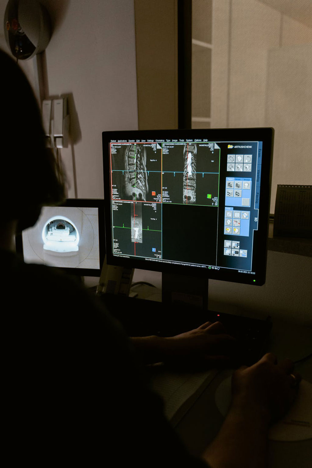
By training ReconGAN on MRI and micro-CT images obtained by taking slice-by-slice pictures of vertebrae acquired from a cadaver, researchers were able to generate realistic micro-structural models of the spine. Using their simulation, the team was also able to virtually enlarge the model, a capability the study says is imperative to understanding and incorporating changes into the entirety of a vertebra’s geometric shape. In this case, the researchers used CT/MRI scans from a 51-year-old female lung cancer patient whose cancer had metastasized to simulate what might happen if cancer weakened some of the vertebrae and how that would affect how much stress the bones could take before fracturing.
The model predicted how much strength parts of the vertebra would lose as a result of the tumors, as well as other changes that could be expected as the cancer progressed. Some of their predictions were confirmed by clinical observations in cancer patients. For a field like orthopedics, using a non-invasive tool like the digital twin can help surgeons understand new therapies, simulate different surgical scenarios and envision how the bone will change over time, either due to bone weakness or to the effects of radiation. The digital twin can also be modified to patient-specific needs, according to the researchers. But this was just a feasibility study and much more work is needed, say the researchers. ReconGAN was trained on data from only one cadaveric sample, and more data is needed for AI to be perfected.
“Spinal fracture increases the risk of patient death by about 15%,” said Soheil Soghrati, co-author of the study and associate professor of mechanical and aerospace engineering at The Ohio State University. “By predicting the outcome of these fractures, our research offers medical experts the opportunity to design better treatment strategies, and help patients make better-informed decisions. What really makes the work in a distinct way is how detailed we were able to model the geometry of the vertebra. We can virtually evolve the same bone from one stage to another.”
“The ultimate goal is to develop a digital twin of everything a surgeon may operate on,” added Soghrati. “Right now, they’re only used for very, very challenging surgeries, but we want to help run those simulations and tune those parameters even more.”
Related Links:
The Ohio State University
Latest General/Advanced Imaging News
- AI-Based Tool Predicts Future Cardiovascular Events in Angina Patients
- AI-Based Tool Accelerates Detection of Kidney Cancer
- New Algorithm Dramatically Speeds Up Stroke Detection Scans
- 3D Scanning Approach Enables Ultra-Precise Brain Surgery
- AI Tool Improves Medical Imaging Process by 90%
- New Ultrasmall, Light-Sensitive Nanoparticles Could Serve as Contrast Agents
- AI Algorithm Accurately Predicts Pancreatic Cancer Metastasis Using Routine CT Images
- Cutting-Edge Angio-CT Solution Offers New Therapeutic Possibilities
- Extending CT Imaging Detects Hidden Blood Clots in Stroke Patients
- Groundbreaking AI Model Accurately Segments Liver Tumors from CT Scans
- New CT-Based Indicator Helps Predict Life-Threatening Postpartum Bleeding Cases
- CT Colonography Beats Stool DNA Testing for Colon Cancer Screening
- First-Of-Its-Kind Wearable Device Offers Revolutionary Alternative to CT Scans
- AI-Based CT Scan Analysis Predicts Early-Stage Kidney Damage Due to Cancer Treatments
- CT-Based Deep Learning-Driven Tool to Enhance Liver Cancer Diagnosis
- AI-Powered Imaging System Improves Lung Cancer Diagnosis
Channels
Radiography
view channel
Routine Mammograms Could Predict Future Cardiovascular Disease in Women
Mammograms are widely used to screen for breast cancer, but they may also contain overlooked clues about cardiovascular health. Calcium deposits in the arteries of the breast signal stiffening blood vessels,... Read more
AI Detects Early Signs of Aging from Chest X-Rays
Chronological age does not always reflect how fast the body is truly aging, and current biological age tests often rely on DNA-based markers that may miss early organ-level decline. Detecting subtle, age-related... Read moreMRI
view channel
MRI Scans Reveal Signature Patterns of Brain Activity to Predict Recovery from TBI
Recovery after traumatic brain injury (TBI) varies widely, with some patients regaining full function while others are left with lasting disabilities. Prognosis is especially difficult to assess in patients... Read more
Novel Imaging Approach to Improve Treatment for Spinal Cord Injuries
Vascular dysfunction in the spinal cord contributes to multiple neurological conditions, including traumatic injuries and degenerative cervical myelopathy, where reduced blood flow can lead to progressive... Read more
AI-Assisted Model Enhances MRI Heart Scans
A cardiac MRI can reveal critical information about the heart’s function and any abnormalities, but traditional scans take 30 to 90 minutes and often suffer from poor image quality due to patient movement.... Read more
AI Model Outperforms Doctors at Identifying Patients Most At-Risk of Cardiac Arrest
Hypertrophic cardiomyopathy is one of the most common inherited heart conditions and a leading cause of sudden cardiac death in young individuals and athletes. While many patients live normal lives, some... Read moreUltrasound
view channel
Wearable Ultrasound Imaging System to Enable Real-Time Disease Monitoring
Chronic conditions such as hypertension and heart failure require close monitoring, yet today’s ultrasound imaging is largely confined to hospitals and short, episodic scans. This reactive model limits... Read more
Ultrasound Technique Visualizes Deep Blood Vessels in 3D Without Contrast Agents
Producing clear 3D images of deep blood vessels has long been difficult without relying on contrast agents, CT scans, or MRI. Standard ultrasound typically provides only 2D cross-sections, limiting clinicians’... Read moreNuclear Medicine
view channel
PET Imaging of Inflammation Predicts Recovery and Guides Therapy After Heart Attack
Acute myocardial infarction can trigger lasting heart damage, yet clinicians still lack reliable tools to identify which patients will regain function and which may develop heart failure.... Read more
Radiotheranostic Approach Detects, Kills and Reprograms Aggressive Cancers
Aggressive cancers such as osteosarcoma and glioblastoma often resist standard therapies, thrive in hostile tumor environments, and recur despite surgery, radiation, or chemotherapy. These tumors also... Read more
New Imaging Solution Improves Survival for Patients with Recurring Prostate Cancer
Detecting recurrent prostate cancer remains one of the most difficult challenges in oncology, as standard imaging methods such as bone scans and CT scans often fail to accurately locate small or early-stage tumors.... Read moreImaging IT
view channel
New Google Cloud Medical Imaging Suite Makes Imaging Healthcare Data More Accessible
Medical imaging is a critical tool used to diagnose patients, and there are billions of medical images scanned globally each year. Imaging data accounts for about 90% of all healthcare data1 and, until... Read more
Global AI in Medical Diagnostics Market to Be Driven by Demand for Image Recognition in Radiology
The global artificial intelligence (AI) in medical diagnostics market is expanding with early disease detection being one of its key applications and image recognition becoming a compelling consumer proposition... Read moreIndustry News
view channel
GE HealthCare and NVIDIA Collaboration to Reimagine Diagnostic Imaging
GE HealthCare (Chicago, IL, USA) has entered into a collaboration with NVIDIA (Santa Clara, CA, USA), expanding the existing relationship between the two companies to focus on pioneering innovation in... Read more
Patient-Specific 3D-Printed Phantoms Transform CT Imaging
New research has highlighted how anatomically precise, patient-specific 3D-printed phantoms are proving to be scalable, cost-effective, and efficient tools in the development of new CT scan algorithms... Read more
Siemens and Sectra Collaborate on Enhancing Radiology Workflows
Siemens Healthineers (Forchheim, Germany) and Sectra (Linköping, Sweden) have entered into a collaboration aimed at enhancing radiologists' diagnostic capabilities and, in turn, improving patient care... Read more


 Guided Devices.jpg)


