Dual-Mode Endoscope Combines Ultrasound and OCT to Offer Unprecedented Insights into Uterine Health
|
By MedImaging International staff writers Posted on 05 Apr 2022 |

Female infertilities are highly associated with poor endometrial receptivity. A receptive endometrium (the lining of the uterus) is generally characterized by the normal uterine cavity, intact endometrial surface, appropriate endometrial thickness, and echo pattern. Acquiring comprehensive structural information is the prerequisite of endometrium assessment, which is beyond the ability of any single-modality imaging method. Researchers have now developed a new endoscope that combines ultrasound with optical coherence tomography (OCT) to assess structural features of the endometrium in unprecedented detail. By providing detailed structural information about the endometrium, the probe could offer a less invasive way to determine if endometrial problems are causing infertility, which affects about 10-20% of women worldwide, as well as help to diagnose other uterine health problems.
The new probe developed by researchers at the Shenzhen Institute of Advanced Technology (Guangdong, China; http://english.siat.cas.cn) could one day help doctors diagnose infertility problems that are related to endometrial receptivity with greater accuracy than current imaging technologies while reducing the need for invasive biopsies. The dual-mode endoscope demonstrated the ability to differentiate between healthy and injured endometrial tissue in rabbit models based on both surface features and depth information. It is the first in vivo demonstration of intrauterine endoscopic imaging in small animals, with a probe measuring just 1.2 mm across.
The endometrium plays a critical role in the ability for a blastocyst to implant in a uterus and grow into a healthy fetus. Failure to implant is recognized as a key bottleneck in the reproductive process, with impaired endometrial receptivity accounting for about two-thirds of implantation failures. The current gold standard method for assessing endometrial receptivity is through biopsies, which require surgically removing and analyzing a small tissue sample. Endoscopic imaging is a less invasive method, but current endoscopes can only identify larger defects in the uterus such as anatomical malformations or polyps, not assess the structure of the endometrium. A vaginal ultrasound can provide information about the thickness of the endometrium and other structural features, but lacks the resolution and contrast needed to comprehensively assess endometrial receptivity.
OCT is an imaging technique that uses relatively long wavelength light (commonly known as near infrared light) to produce high-resolution images from within scattering media. It has been adapted for diagnostic tools in several medical fields including ophthalmology, cardiology and dermatology. Previous studies have shown OCT imaging can be used to identify structural features of the endometrium that are associated with implantation failures. For the new study, researchers improved upon a prototype they had previously developed to combine OCT and ultrasound imaging in a single probe. The OCT modality provides detailed information about the superficial endometrium including its surface information, while ultrasound provides insights about its full thickness. Since multiple features of the endometrium affect implantation success, combining these imaging modalities provides a more accurate picture of endometrial receptivity than either mode individually.
The catheter is designed to pass through the cervix, enter the uterine cavity and inject water to facilitate high-resolution imaging. A series of tiny custom-designed optical and ultrasonic components are arranged within the catheter to achieve both ultrasound and OCT mode. The improved probe also uses a single-mode fiber, which offers higher resolution and reduced noise for the OCT mode. In addition, the researchers used a metal coil to allow the probe to rotate for a 360-degree full-field of view once it is inside the uterus. To test the endoscope, the researchers used it to image the uterine lining of four anesthetized rabbits. Some of the rabbits were healthy while others had undergone a procedure to wash the endometrium with ethanol for different lengths of time, damaging the tissue to varying degrees.
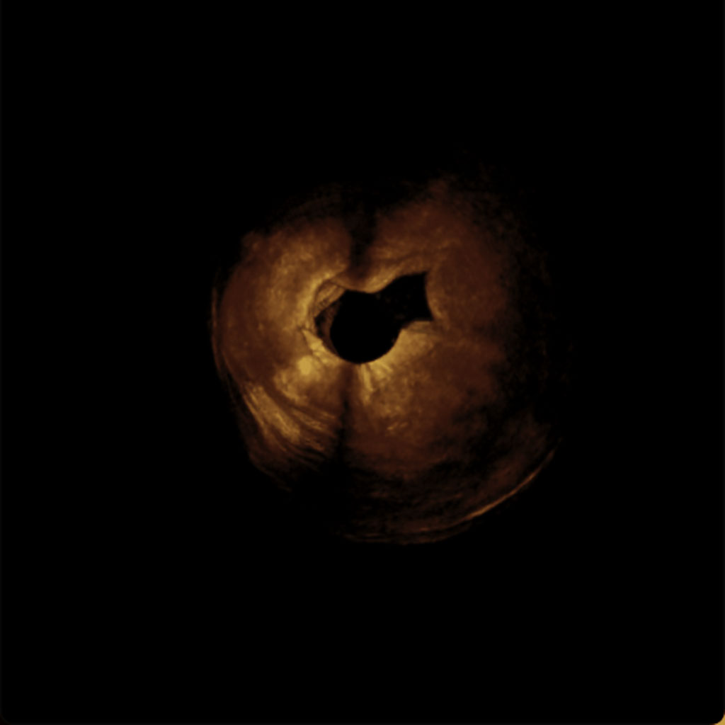
The researchers quantified features of the endometrium including its thickness, distribution and surface roughness separately in ultrasonic and OCT modalities. The OCT images showed that healthy endometrial tissues had a smoother and more continuous surface, while damaged tissues were more rough. In ultrasound images, the endometrium was found to be thicker in healthy tissues and thinner in areas that had been damaged. While each modality provided valuable information on its own, it wasn’t until the researchers combined the information from each that they were able to comprehensively and accurately evaluate the degree of tissue injury.
The probe also provided echo patterns that were similar to what can be obtained with vaginal ultrasound but with better resolution. In addition, the images revealed physical features such as polyp-like formations as small as 200 microns, demonstrating the probe’s ability to discern tiny lesions that could affect endometrial health. The researchers plan to add a photoacoustic mode to increase the probe’s ability to observe blood flow and information about the vascular networks in the uterine lining. In addition, they are working to improve the size, resolution and imaging range of the imaging catheter to make it more practical for clinical use in humans.
“This tool combines the two techniques of ultrasound and OCT, allowing it to obtain more information and provide a more accurate assessment of endometrial status than traditional vaginal ultrasound,” said research team leader Xiaojing Gong from the Shenzhen Institutes of Advanced Technology of the Chinese Academy of Sciences. “It has the potential to be used for basic endometrial research and to further advance clinical assessment of endometrial receptivity and other endometrial-related diseases.”
“The system can obtain the thickness information of the endometrium, the echo pattern of the endometrium and information about damage to the endometrial surface, which play an important role in the evaluation of endometrial receptivity,” added Gong. “It also has the potential to detect diseases in the uterus, such as endometrial cancer and uterine fibroids.”
Related Links:
Shenzhen Institute of Advanced Technology
Latest Ultrasound News
- Tiny Magnetic Robot Takes 3D Scans from Deep Within Body
- High Resolution Ultrasound Speeds Up Prostate Cancer Diagnosis
- World's First Wireless, Handheld, Whole-Body Ultrasound with Single PZT Transducer Makes Imaging More Accessible
- Artificial Intelligence Detects Undiagnosed Liver Disease from Echocardiograms
- Ultrasound Imaging Non-Invasively Tracks Tumor Response to Radiation and Immunotherapy
- AI Improves Detection of Congenital Heart Defects on Routine Prenatal Ultrasounds
- AI Diagnoses Lung Diseases from Ultrasound Videos with 96.57% Accuracy
- New Contrast Agent for Ultrasound Imaging Ensures Affordable and Safer Medical Diagnostics
- Ultrasound-Directed Microbubbles Boost Immune Response Against Tumors
- POC Ultrasound Enhances Early Pregnancy Care and Cuts Emergency Visits
- AI-Based Models Outperform Human Experts at Identifying Ovarian Cancer in Ultrasound Images
- Automated Breast Ultrasound Provides Alternative to Mammography in Low-Resource Settings
- Transparent Ultrasound Transducer for Photoacoustic and Ultrasound Endoscopy to Improve Diagnostic Accuracy
- Wearable Ultrasound Patch Enables Continuous Blood Pressure Monitoring
- AI Image-Recognition Program Reads Echocardiograms Faster, Cuts Results Wait Time
- Ultrasound Device Non-Invasively Improves Blood Circulation in Lower Limbs
Channels
Radiography
view channel
AI-Powered Imaging Technique Shows Promise in Evaluating Patients for PCI
Percutaneous coronary intervention (PCI), also known as coronary angioplasty, is a minimally invasive procedure where small metal tubes called stents are inserted into partially blocked coronary arteries... Read more
Higher Chest X-Ray Usage Catches Lung Cancer Earlier and Improves Survival
Lung cancer continues to be the leading cause of cancer-related deaths worldwide. While advanced technologies like CT scanners play a crucial role in detecting lung cancer, more accessible and affordable... Read moreMRI
view channel
Ultra-Powerful MRI Scans Enable Life-Changing Surgery in Treatment-Resistant Epileptic Patients
Approximately 360,000 individuals in the UK suffer from focal epilepsy, a condition in which seizures spread from one part of the brain. Around a third of these patients experience persistent seizures... Read more
AI-Powered MRI Technology Improves Parkinson’s Diagnoses
Current research shows that the accuracy of diagnosing Parkinson’s disease typically ranges from 55% to 78% within the first five years of assessment. This is partly due to the similarities shared by Parkinson’s... Read more
Biparametric MRI Combined with AI Enhances Detection of Clinically Significant Prostate Cancer
Artificial intelligence (AI) technologies are transforming the way medical images are analyzed, offering unprecedented capabilities in quantitatively extracting features that go beyond traditional visual... Read more
First-Of-Its-Kind AI-Driven Brain Imaging Platform to Better Guide Stroke Treatment Options
Each year, approximately 800,000 people in the U.S. experience strokes, with marginalized and minoritized groups being disproportionately affected. Strokes vary in terms of size and location within the... Read moreNuclear Medicine
view channel
Novel PET Imaging Approach Offers Never-Before-Seen View of Neuroinflammation
COX-2, an enzyme that plays a key role in brain inflammation, can be significantly upregulated by inflammatory stimuli and neuroexcitation. Researchers suggest that COX-2 density in the brain could serve... Read more
Novel Radiotracer Identifies Biomarker for Triple-Negative Breast Cancer
Triple-negative breast cancer (TNBC), which represents 15-20% of all breast cancer cases, is one of the most aggressive subtypes, with a five-year survival rate of about 40%. Due to its significant heterogeneity... Read moreGeneral/Advanced Imaging
view channel
AI-Powered Imaging System Improves Lung Cancer Diagnosis
Given the need to detect lung cancer at earlier stages, there is an increasing need for a definitive diagnostic pathway for patients with suspicious pulmonary nodules. However, obtaining tissue samples... Read more
AI Model Significantly Enhances Low-Dose CT Capabilities
Lung cancer remains one of the most challenging diseases, making early diagnosis vital for effective treatment. Fortunately, advancements in artificial intelligence (AI) are revolutionizing lung cancer... Read moreImaging IT
view channel
New Google Cloud Medical Imaging Suite Makes Imaging Healthcare Data More Accessible
Medical imaging is a critical tool used to diagnose patients, and there are billions of medical images scanned globally each year. Imaging data accounts for about 90% of all healthcare data1 and, until... Read more
Global AI in Medical Diagnostics Market to Be Driven by Demand for Image Recognition in Radiology
The global artificial intelligence (AI) in medical diagnostics market is expanding with early disease detection being one of its key applications and image recognition becoming a compelling consumer proposition... Read moreIndustry News
view channel
GE HealthCare and NVIDIA Collaboration to Reimagine Diagnostic Imaging
GE HealthCare (Chicago, IL, USA) has entered into a collaboration with NVIDIA (Santa Clara, CA, USA), expanding the existing relationship between the two companies to focus on pioneering innovation in... Read more
Patient-Specific 3D-Printed Phantoms Transform CT Imaging
New research has highlighted how anatomically precise, patient-specific 3D-printed phantoms are proving to be scalable, cost-effective, and efficient tools in the development of new CT scan algorithms... Read more
Siemens and Sectra Collaborate on Enhancing Radiology Workflows
Siemens Healthineers (Forchheim, Germany) and Sectra (Linköping, Sweden) have entered into a collaboration aimed at enhancing radiologists' diagnostic capabilities and, in turn, improving patient care... Read more


