MRI Post-Processing Platform Visualizes Entire Brain in Minutes
|
By MedImaging International staff writers Posted on 29 Aug 2021 |

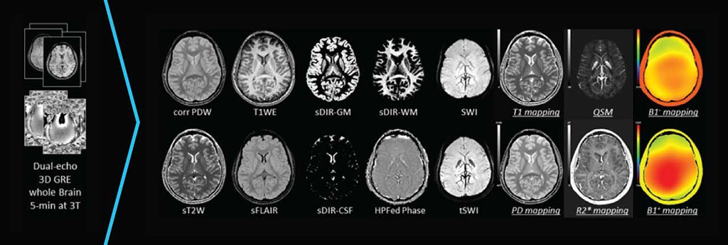
Image: STAGE’s 10 qualitative and six quantitative image outputs (Photo courtesy of SpinTech)
A new magnetic resonance imaging (MRI) software platform enables comprehensive, quantitative brain imaging with enhanced visualization in significantly less time than conventional approaches.
The SpinTech (Holland, MI, USA) STAGE platform is a powerful, rapid, multi-contrast imaging method that allows scanning the entire brain in roughly five minutes. STAGE allows acquisition of 16 brain imaging contrasts, including maps of Tl, R2*, and proton density (PD), enhanced Tl weighted images, susceptibility weighted imaging (SWI) images, susceptibility weighted image map (SWIM) images, pseudo-SWIM (pSWIM) images, modified pSWIM (mpSWIM) images, true SWI (tSWI) images, MR angiography images, simulated dual-inversion recovery (DIR) images.
The STAGE system consists of a dedicated computer connected to the user’s local area network. The computer receives DICOM data from a MRI scan using a specific 3D GRE scan protocol (the STAGE protocol), and outputs in return numerous DICOM datasets with different types of contrast to the picture archiving and communication systems (PACS) server. STAGE is compatible with both 1.5T and 3T systems across manufacturers, and provides standardized outputs that can enable more reliable longitudinal comparison across different MRI machines.
“STAGE's novel acquisition technique and post-processing software, which grew from the world's top MRI research groups, is designed to enhance visualization of biomarkers that couldn't be seen in the brain before while improving throughput and accuracy, making it a significant advancement in imaging,” said Ward Detweiler, President and CEO of SpinTech. “We are incredibly excited to make this game-changing technology available for clinical use in hospitals and imaging centers.”
“Radiologists have long struggled to obtain comprehensive, high-quality clinical data within very constricted scanning windows,” said Mark Haacke, PhD, founder and CSO of SpinTech. “Now, they don't need to choose which sequences to run, just what they are going to examine, as all the data they need has already been collected. On top of the enhanced imaging data, radiology groups will also benefit from increased patient throughput. And, of course, patients experience shorter scan times, so everybody wins.”
Related Links:
SpinTech
Latest MRI News
- Novel Imaging Approach to Improve Treatment for Spinal Cord Injuries
- AI-Assisted Model Enhances MRI Heart Scans
- AI Model Outperforms Doctors at Identifying Patients Most At-Risk of Cardiac Arrest
- New MRI Technique Reveals Hidden Heart Issues
- Shorter MRI Exam Effectively Detects Cancer in Dense Breasts
- MRI to Replace Painful Spinal Tap for Faster MS Diagnosis
- MRI Scans Can Identify Cardiovascular Disease Ten Years in Advance
- Simple Brain Scan Diagnoses Parkinson's Disease Years Before It Becomes Untreatable
- Cutting-Edge MRI Technology to Revolutionize Diagnosis of Common Heart Problem
- New MRI Technique Reveals True Heart Age to Prevent Attacks and Strokes
- AI Tool Predicts Relapse of Pediatric Brain Cancer from Brain MRI Scans
- AI Tool Tracks Effectiveness of Multiple Sclerosis Treatments Using Brain MRI Scans
- Ultra-Powerful MRI Scans Enable Life-Changing Surgery in Treatment-Resistant Epileptic Patients
- AI-Powered MRI Technology Improves Parkinson’s Diagnoses
- Biparametric MRI Combined with AI Enhances Detection of Clinically Significant Prostate Cancer
- First-Of-Its-Kind AI-Driven Brain Imaging Platform to Better Guide Stroke Treatment Options
Channels
Radiography
view channel
X-Ray Breakthrough Captures Three Image-Contrast Types in Single Shot
Detecting early-stage cancer or subtle changes deep inside tissues has long challenged conventional X-ray systems, which rely only on how structures absorb radiation. This limitation keeps many microstructural... Read more
AI Generates Future Knee X-Rays to Predict Osteoarthritis Progression Risk
Osteoarthritis, a degenerative joint disease affecting over 500 million people worldwide, is the leading cause of disability among older adults. Current diagnostic tools allow doctors to assess damage... Read moreUltrasound
view channel
Wearable Ultrasound Imaging System to Enable Real-Time Disease Monitoring
Chronic conditions such as hypertension and heart failure require close monitoring, yet today’s ultrasound imaging is largely confined to hospitals and short, episodic scans. This reactive model limits... Read more
Ultrasound Technique Visualizes Deep Blood Vessels in 3D Without Contrast Agents
Producing clear 3D images of deep blood vessels has long been difficult without relying on contrast agents, CT scans, or MRI. Standard ultrasound typically provides only 2D cross-sections, limiting clinicians’... Read moreNuclear Medicine
view channel
PET Imaging of Inflammation Predicts Recovery and Guides Therapy After Heart Attack
Acute myocardial infarction can trigger lasting heart damage, yet clinicians still lack reliable tools to identify which patients will regain function and which may develop heart failure.... Read more
Radiotheranostic Approach Detects, Kills and Reprograms Aggressive Cancers
Aggressive cancers such as osteosarcoma and glioblastoma often resist standard therapies, thrive in hostile tumor environments, and recur despite surgery, radiation, or chemotherapy. These tumors also... Read more
New Imaging Solution Improves Survival for Patients with Recurring Prostate Cancer
Detecting recurrent prostate cancer remains one of the most difficult challenges in oncology, as standard imaging methods such as bone scans and CT scans often fail to accurately locate small or early-stage tumors.... Read moreGeneral/Advanced Imaging
view channel
3D Scanning Approach Enables Ultra-Precise Brain Surgery
Precise navigation is critical in neurosurgery, yet even small alignment errors can affect outcomes when operating deep within the brain. A new 3D surface-scanning approach now provides a radiation-free... Read more
AI Tool Improves Medical Imaging Process by 90%
Accurately labeling different regions within medical scans, a process known as medical image segmentation, is critical for diagnosis, surgery planning, and research. Traditionally, this has been a manual... Read more
New Ultrasmall, Light-Sensitive Nanoparticles Could Serve as Contrast Agents
Medical imaging technologies face ongoing challenges in capturing accurate, detailed views of internal processes, especially in conditions like cancer, where tracking disease development and treatment... Read more
AI Algorithm Accurately Predicts Pancreatic Cancer Metastasis Using Routine CT Images
In pancreatic cancer, detecting whether the disease has spread to other organs is critical for determining whether surgery is appropriate. If metastasis is present, surgery is not recommended, yet current... Read moreImaging IT
view channel
New Google Cloud Medical Imaging Suite Makes Imaging Healthcare Data More Accessible
Medical imaging is a critical tool used to diagnose patients, and there are billions of medical images scanned globally each year. Imaging data accounts for about 90% of all healthcare data1 and, until... Read more
Global AI in Medical Diagnostics Market to Be Driven by Demand for Image Recognition in Radiology
The global artificial intelligence (AI) in medical diagnostics market is expanding with early disease detection being one of its key applications and image recognition becoming a compelling consumer proposition... Read moreIndustry News
view channel
GE HealthCare and NVIDIA Collaboration to Reimagine Diagnostic Imaging
GE HealthCare (Chicago, IL, USA) has entered into a collaboration with NVIDIA (Santa Clara, CA, USA), expanding the existing relationship between the two companies to focus on pioneering innovation in... Read more
Patient-Specific 3D-Printed Phantoms Transform CT Imaging
New research has highlighted how anatomically precise, patient-specific 3D-printed phantoms are proving to be scalable, cost-effective, and efficient tools in the development of new CT scan algorithms... Read more
Siemens and Sectra Collaborate on Enhancing Radiology Workflows
Siemens Healthineers (Forchheim, Germany) and Sectra (Linköping, Sweden) have entered into a collaboration aimed at enhancing radiologists' diagnostic capabilities and, in turn, improving patient care... Read more


