AI Algorithm Reduces Unnecessary Radiation Exposure in Traumatic Neuroradiological CT Scans
Posted on 25 Sep 2024
Traumatic neuroradiological emergencies encompass conditions that require immediate and accurate diagnosis for effective treatment and optimal patient outcomes. These emergencies can include injuries to the brain or spinal cord. Computed tomography (CT) is one of the most widely used imaging techniques in such situations due to its availability and speed, making it a vital tool in emergency departments for assessing the severity of injuries and guiding treatment. However, a significant limitation of CT scans is the relationship between radiation dose and image quality. CT scans involve exposure to ionizing radiation, which poses a potential long-term risk to patients, including the development of cancer. Recently, modern artificial intelligence (AI) reconstruction algorithms have emerged, offering the potential to reduce radiation doses while maintaining high image quality. These AI algorithms could minimize the risks associated with ionizing radiation and improve patient outcomes, though the extent to which they reduce radiation exposure in traumatic neuroradiological CT scans has not been extensively studied.
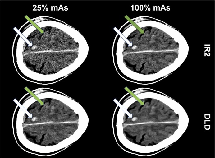
To address this, researchers at Eberhard Karls-University Tuebingen (Tuebingen, Germany) conducted a comparative study to evaluate the performance of a deep learning-based denoising (DLD) algorithm in CT scans of patients with traumatic neuroradiological emergencies. They proposed that the use of these algorithms could allow for high-quality imaging at reduced radiation doses, thereby improving patient care by minimizing unnecessary radiation exposure. The retrospective, single-center study involved 100 patients who had undergone neuroradiological trauma CT scans. Both full-dose (100%) and low-dose (25%) simulated scans were processed using iterative reconstruction (IR2) and the DLD algorithm. Four neuroradiologists assessed the subjective and objective quality of the images, alongside a clinical endpoint analysis. Bayesian sensitivity and specificity were calculated with 95% credible intervals.

The study found that the DLD algorithm produced high-quality, fully diagnostic CT images at just 25% of the standard radiation dose, demonstrating its potential to enhance patient care by reducing unnecessary radiation exposure. The algorithm’s ability to maintain image quality at significantly lower radiation doses highlights its promise in addressing concerns about radiation exposure, particularly in the context of frequent head CT use in neuroradiological emergencies. This issue has been a growing concern for both patients and physicians, particularly regarding the carcinogenic risks associated with long-term radiation exposure, especially in younger patients. The findings of this study contribute to ongoing efforts to reduce radiation doses in medical imaging and emphasize the importance of further research into dose-reduction techniques.
Related Links:
Eberhard Karls-University Tuebingen


