Compact MRI Addresses Neonatal ICU Imaging Challenges
|
By MedImaging International staff writers Posted on 24 Dec 2020 |


|
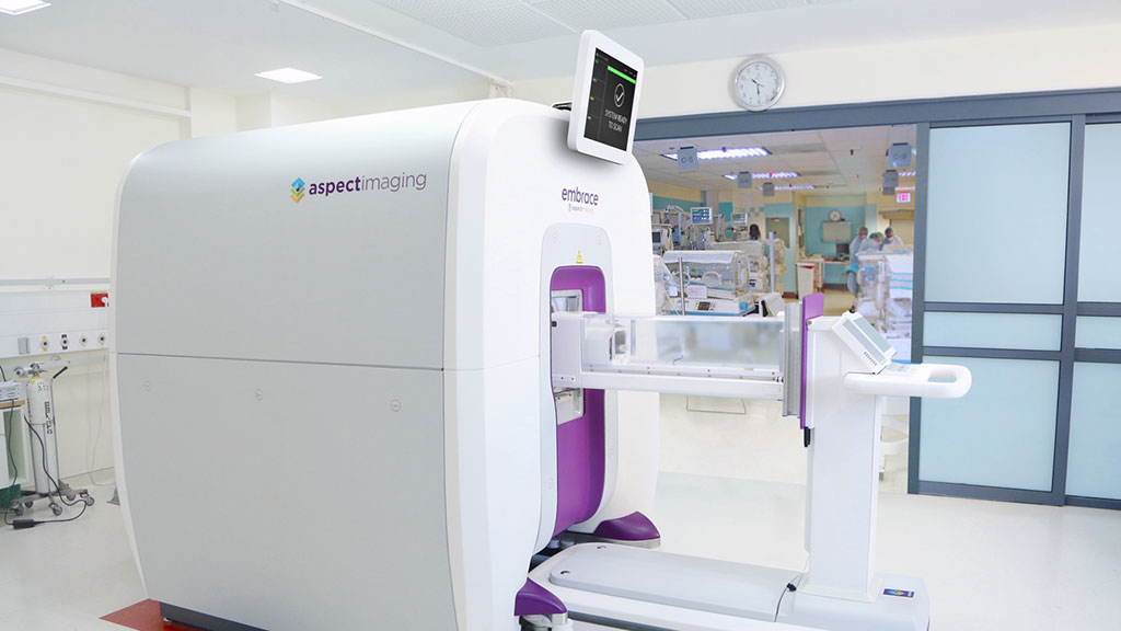
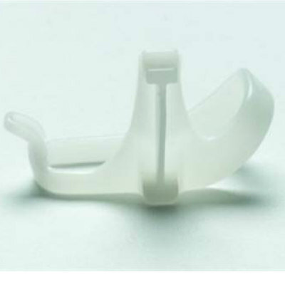
Image: The Embrace Neonatal MRI System (Photo courtesy of Aspect Imaging)
An advanced magnetic resonance imaging (MRI) system performs scans in the neonatal intensive care unit (nICU) itself, eliminating transportation of the fragile patients to radiology departments.
The Aspect Imaging (Shoham, Israel) Embrace Neonatal MRI System is a fully enclosed system for babies weighing between one and 4.5 kilograms and a head circumference of up to 38 centimeters. Based on the proprietary magnetic technology, which does not require cooling, the self-shielded system eliminates the need for a safety zone or radiofrequency (RF)-shielded room, enabling installation inside a nICU. Medical teams can thus assure the quality care for babies before, during, and after the procedure.
Neonates are placed inside a self-contained incubator bed which minimizes movement while providing a temperature-controlled (36.5o Celsius) environment and continuous monitoring of vital signs. System features include a workflow that allows prep and scan in less than one hour; a dedicated head coil for diagnostic quality brain imaging; three different swaddles for restricting movement and routing of tubing and monitoring leads; and PACS/HIS/RIS connectivity with DICOM compatibility. Embrace also reduces parental emotional anxiety by allowing them to view their baby on the full-color video display during the scanning procedure.
“Aspect Imaging supports neonatologists and neuroradiologists in making accurate diagnostic decisions by delivering clinical information to them easily via connected picture archiving and communication system servers, without the disruption of transporting babies out of the NICU,” said Ashish Kaul, a senior research analyst at Frost and Sullivan (Mountain View, CA, USA). “The company aims to disrupt and redefine the entire neonatal medical imaging space and has shown strong growth potential across geographic regions.”





The Aspect Imaging (Shoham, Israel) Embrace Neonatal MRI System is a fully enclosed system for babies weighing between one and 4.5 kilograms and a head circumference of up to 38 centimeters. Based on the proprietary magnetic technology, which does not require cooling, the self-shielded system eliminates the need for a safety zone or radiofrequency (RF)-shielded room, enabling installation inside a nICU. Medical teams can thus assure the quality care for babies before, during, and after the procedure.
Neonates are placed inside a self-contained incubator bed which minimizes movement while providing a temperature-controlled (36.5o Celsius) environment and continuous monitoring of vital signs. System features include a workflow that allows prep and scan in less than one hour; a dedicated head coil for diagnostic quality brain imaging; three different swaddles for restricting movement and routing of tubing and monitoring leads; and PACS/HIS/RIS connectivity with DICOM compatibility. Embrace also reduces parental emotional anxiety by allowing them to view their baby on the full-color video display during the scanning procedure.
“Aspect Imaging supports neonatologists and neuroradiologists in making accurate diagnostic decisions by delivering clinical information to them easily via connected picture archiving and communication system servers, without the disruption of transporting babies out of the NICU,” said Ashish Kaul, a senior research analyst at Frost and Sullivan (Mountain View, CA, USA). “The company aims to disrupt and redefine the entire neonatal medical imaging space and has shown strong growth potential across geographic regions.”

Breast Localization System
MAMMOREP LOOP
Mammo DR Retrofit Solution
DR Retrofit Mammography
X-Ray Illuminator
X-Ray Viewbox Illuminators
Ultrasound Needle Guidance System
SonoSite L25
Latest MRI News
- Cardiac MRI Detects Hidden Heart Dysfunction After Heart Attack
- AI Tool Reveals Cortical Lesions on Standard MRI in Multiple Sclerosis
- AI Reconstruction Tool Speeds Dynamic Breast MRI and Improves Cancer Detection
- International Study Assesses AI for Prostate Cancer MRI Interpretation
- AI Approach Could Shorten Advanced Brain MRI Scans by Up to 90%
- Cardiac MRI Measure Improves Risk Prediction in Tricuspid Regurgitation
- AI System Improves Accuracy of Cardiac MRI Interpretation
- Deep Learning Model Predicts Alzheimer’s Disease Outcomes from Baseline MRI
- Blood-Brain Barrier Imaging Adds Risk Insight to Standard Stroke MRI
- AI Body Composition MRI Analysis Predicts Cardiometabolic Disease Risk
- AI MRI Tool Quantifies Muscle Fat to Assess Cardiometabolic Risk
- Advanced MRI Visualizes CSF Motion Changes After Mild Traumatic Brain Injury
- MRI Tool Enables Long-Term Tracking of Transplanted Cardiac Cells
- MRI-Based AI Tool Supports Differentiation of Parkinsonian Syndromes
- MRI-Derived Biomarker Improves Risk Stratification in Glioblastoma
- Combined Imaging Approach Identifies Cause of Heart Attack without Coronary Blockage
Channels
Radiography
view channel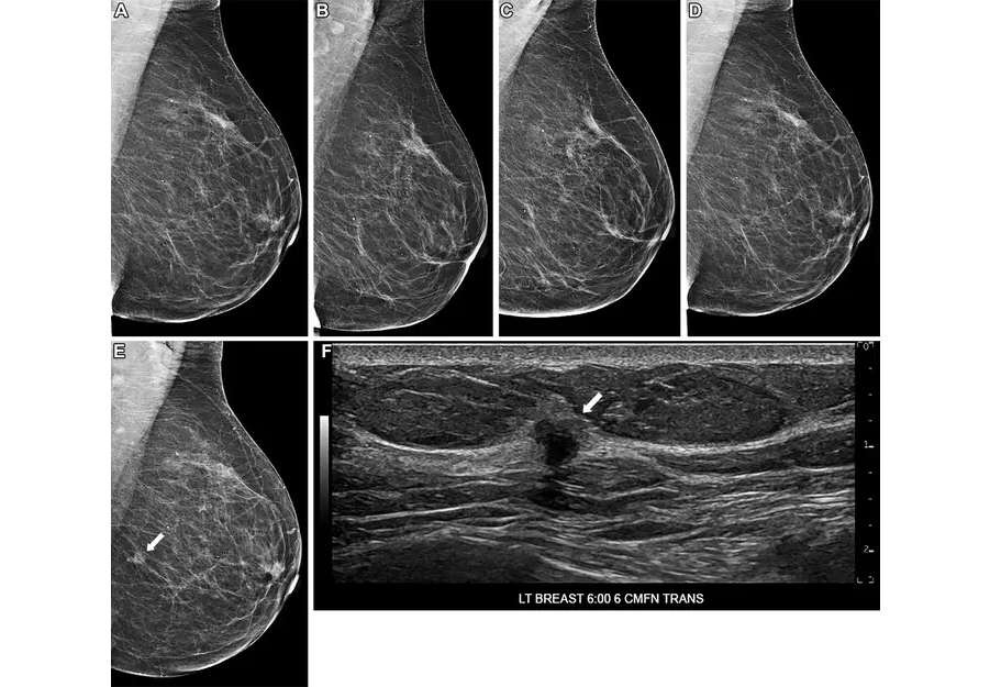
AI Tool Predicts Five-Year Breast Cancer Risk from Mammograms
Breast cancer risk assessment during routine screening is difficult because many women who develop the disease have no known genetic mutations or family history. Static risk tools provide limited discrimination... Read more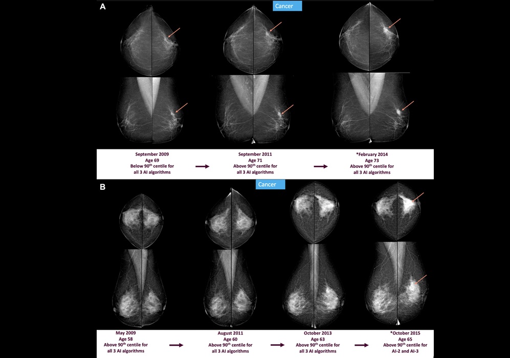
AI Mammography Tools Detect Early Breast Cancer Signs Years Before Diagnosis
Breast cancer screening aims to detect tumors before symptoms develop, but subtle mammographic changes can appear years before diagnosis and may be missed during routine reads. Delayed detection can lead... Read more
Ultrasound
view channel
Simple 2D Ultrasound Tool Accurately Estimates Placental Volume
Stillbirth remains a major cause of perinatal loss, with about 21,000 cases annually in the United States. Although placental abnormalities are linked to adverse pregnancy outcomes, routine monitoring... Read more
Portable 3D Ultrasound System Enables Reproducible Breast Cancer Monitoring
Breast cancer can develop between annual mammograms, and these interval cancers account for 20% to 30% of cases and tend to be more aggressive. The challenge is pronounced in people with dense breast tissue.... Read more
Nuclear Medicine
view channel
Radiotherapy Professionals Embrace AI as a Clinical Support Tool
Radiotherapy planning requires precise identification of organs at risk near a tumor, but manual contouring is repetitive and time-consuming, often delaying plan completion and straining specialist capacity.... Read more
Second PSMA PET Identifies Disease in More Than Half of Negative Cases
Biochemical recurrence of prostate cancer after prostatectomy or radiation can be difficult to localize when imaging is negative. Uncertain disease sites may delay salvage therapy and expose patients to... Read more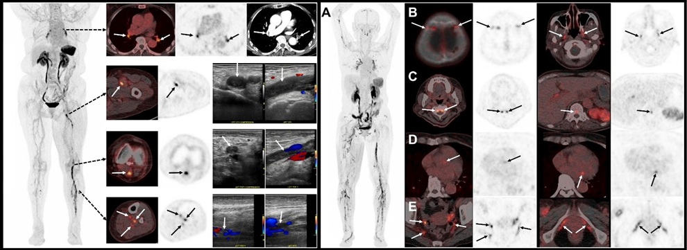
![Image: (A-C) Representative whole-body maximum-intensity projection images and regional fused PET/CT images from three histologically confirmed osteosarcoma patients who underwent paired [68Ga]Ga-B7-H3-BCH PET/CT and 18F-FDGE PET/CT within 7 days. (D) Multimodal imaging evaluation of patient 3, including X-ray, MRI (T2-weighted imaging, T2WI), CT, and B7-H3 PET/CT. (Photo courtesy of SNMMI)](https://globetechcdn.com/mobile_medicalimaging/images/stories/articles/article_images/2026-06-07/294811110_image_1.jpg "Image: (A-C) Representative whole-body maximum-intensity projection images and regional fused PET/CT images from three histologically confirmed osteosarcoma patients who underwent paired [68Ga]Ga-B7-H3-BCH PET/CT and 18F-FDGE PET/CT within 7 days. (D) Multimodal imaging evaluation of patient 3, including X-ray, MRI (T2-weighted imaging, T2WI), CT, and B7-H3 PET/CT. (Photo courtesy of SNMMI)")
General/Advanced Imaging
view channel
Virtual Staining Technique Creates Histology Images from CT Data
Pulmonary hypertension, a disorder marked by pathological remodeling of the pulmonary vessels, often requires detailed histologic assessment. Yet routine pathology remains anchored in labor‑intensive,... Read more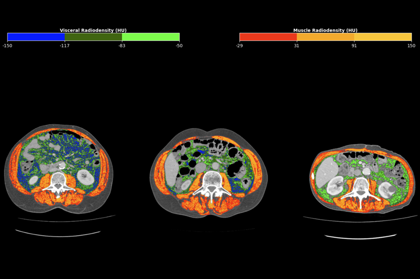
CT-Derived Biomarker Predicts Outcomes in Gastric Cancer
Gastric cancer, also known as stomach cancer, is the fifth most common malignancy worldwide and often shows heterogeneous outcomes even within the same stage. Prognostic estimates typically rely on tumor-centric... Read more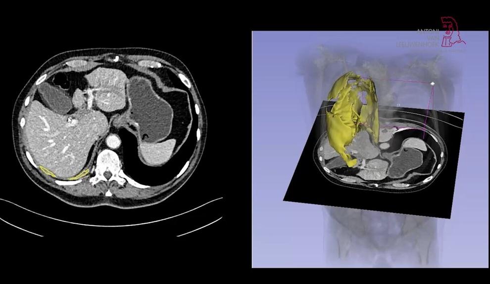
AI Tool Enhances Response Assessment and Survival Prediction in Pleural Mesothelioma
Pleural mesothelioma, a cancer that grows as a thin, irregular layer along the lung wall, is difficult to measure on imaging. Clinicians rely on diameter-based Response Evaluation Criteria in Solid Tumors... Read more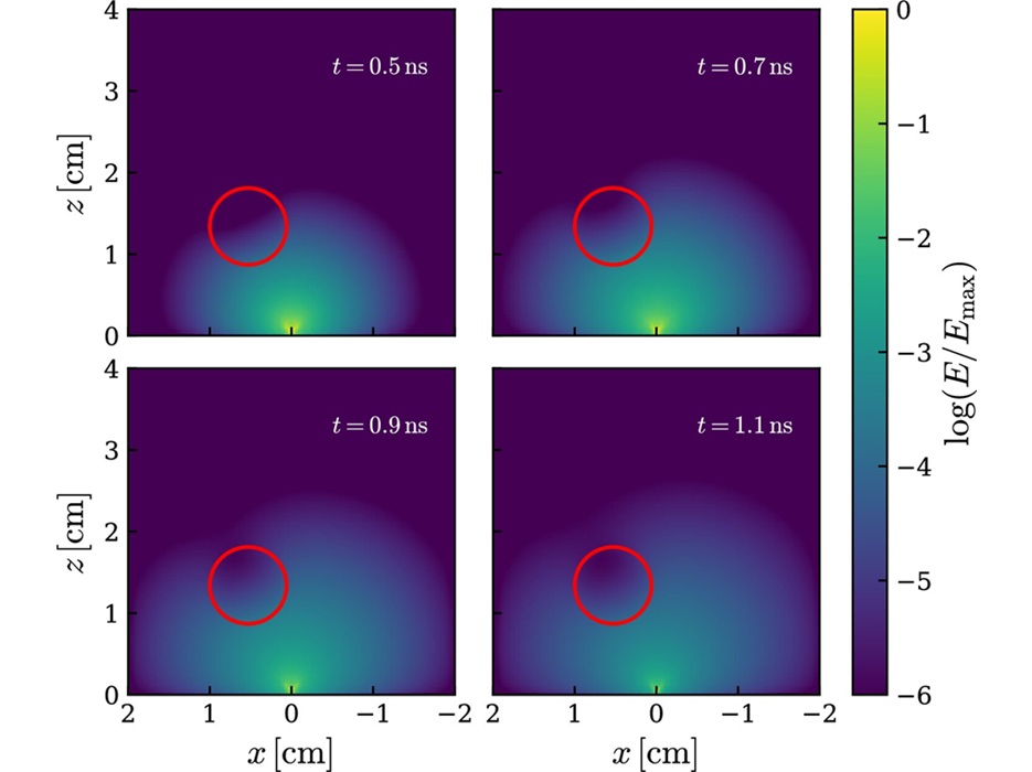
Imaging IT
view channel
Ambient AI Reporting Platform Streamlines Radiology Reporting
Radiology departments face growing imaging volumes and staffing shortages, creating reporting bottlenecks and pressure to maintain turnaround times. Conventional dictation tools document findings after... Read more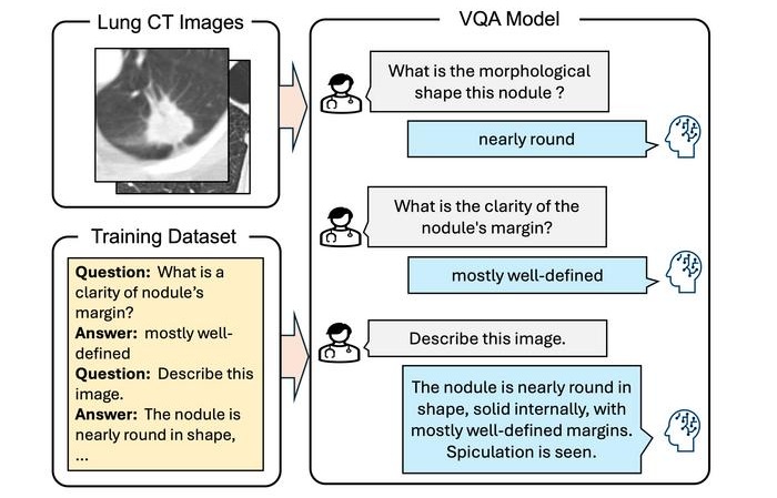
Interactive AI Tool Supports Explainable Lung Nodule Assessment
Lung cancer is a leading cause of cancer mortality, and timely characterization of pulmonary nodules on chest computed tomography (CT) is essential for directing care. Interpreting nodule morphology demands... Read more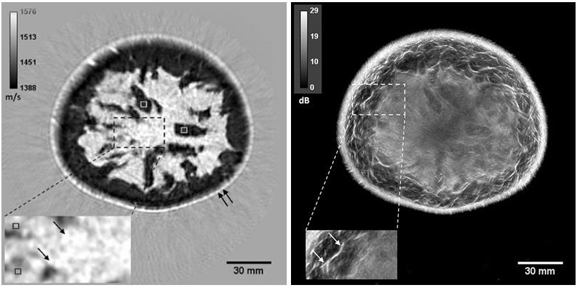
Breast Imaging Software Enhances Visualization and Tissue Characterization in Challenging Cases
Breast imaging can be particularly challenging in cases involving small breasts or implants, where image reconstruction and tissue characterization may be limited. Clinicians also need reproducible analysis... Read more
Industry News
view channel
GE HealthCare Showcases AI-Enabled Nuclear Medicine Portfolio at SNMMI 2026
Nuclear medicine is expanding rapidly as health systems adopt theranostics and broaden access to radiopharmaceuticals, increasing demand for scalable operations and consistent diagnostic confidence.... Read more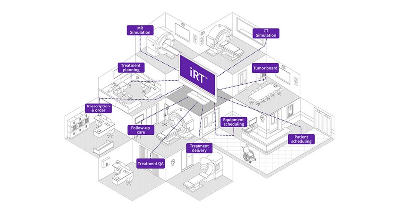
GE HealthCare Highlights AI-Supported Radiation Therapy Tools at ESTRO 2026
At the European Society for Radiotherapy and Oncology (ESTRO) 2026 Congress in Stockholm, GE HealthCare is highlighting Intelligent Radiation Therapy (iRT), MIM Software innovations, and BK Medical surgical... Read more