AI Approach Could Shorten Advanced Brain MRI Scans by Up to 90%
Posted on 28 May 2026
Long acquisition times for advanced brain magnetic resonance imaging (MRI) can limit access, extend waiting lists, and disrupt clinical workflows. Reducing data requirements without sacrificing image fidelity remains a persistent technical barrier. Faster protocols are especially relevant for patients who cannot tolerate lengthy scans. To help address this challenge, investigators have developed a simulation- and artificial intelligence–based approach that reconstructs brain microstructural information using far less data.
Researchers at the Institute for Neurosciences (IN), a joint center of the Spanish National Research Council (CSIC) and Miguel Hernández University of Elche (UMH; Elche, Spain), report a strategy that trains neural networks entirely on computer-simulated diffusion signals. The method is designed to estimate model parameters that function as tissue biomarkers from a very small number of diffusion-weighted images. The work targets brain MRI and aims to increase the clinical information available from shorter scans.
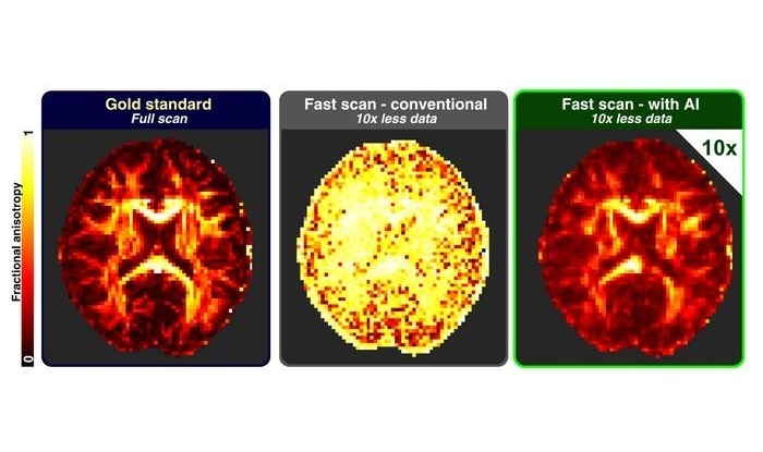
Instead of relying on large clinical datasets, the approach uses a physics-based model of water diffusion in brain tissue to generate training data. Artificial intelligence then reconstructs detailed microstructural features from the resulting signals. By decoupling training from patient availability, the method seeks to minimize dataset bias and avoid privacy constraints.
The study indicates that certain advanced MRI scans could be shortened by up to 90% while preserving high accuracy. The network achieved a very high level of accuracy using only 10% of the usual diffusion data. In practical terms, the time to obtain equivalent information could drop from about 40 minutes to roughly 8, which could help hospitals address long waiting lists.
The study was published in Communications Medicine. The approach also opens possibilities for earlier investigation of neurodegenerative diseases such as Alzheimer’s disease, which can have a preclinical phase lasting up to two decades. The system further enables reanalysis of legacy MRI datasets to extract new information about neurological disease.
“Reducing the acquisition time required makes it possible to incorporate much more advanced MRI techniques, resulting in a greater amount of clinical information available to medical staff,” said Silvia De Santis, who leads the Translational Imaging Biomarkers Laboratory at the IN CSIC-UMH.
“We have shown that our network, trained entirely on simulations, can achieve a very high level of accuracy using only 10% of the data. This could have a direct impact in clinical settings, especially in hospitals with very long waiting lists,” said Maximilian Eggl, who leads the AI-inspired Biomarkers of Brain Structure and Function research line at the IN CSIC-UMH.
Related Links:
Miguel Hernández University