Fujifilm Previews World's First Glass-Free Digital Radiography Detector at RSNA 2019 Image
|
By MedImaging International staff writers Posted on 02 Dec 2019 |

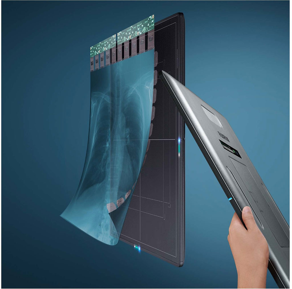
Image: Glass-Free Digital Radiography Detector (Photo courtesy of FUJIFILM Medical Systems U.S.A., Inc.)
FUJIFILM Medical Systems U.S.A., Inc. (Lexington, MA, USA) previewed its new FDR D-EVO III DR detector at the 105th scientific assembly and annual meeting of the Radiological Society of North America (RSNA) held from December 1 – 6, 2019 in Chicago, USA. FUJIFILM Medical Systems, which is part of the FUJIFILM Healthcare portfolio, is a provider of diagnostic imaging products and medical informatics solutions ranging from digital X-ray systems (DR: detectors, mobiles, rooms and 3D mammography), to the comprehensive Synapse Enterprise Imaging portfolio, to full-field digital mammography systems with digital breast tomosynthesis, and computed tomography solutions.
The FDR D-EVO III previewed at RSNA 2019 is FUJIFILM's third-generation digital X-ray detector featuring a sleek, thin design and status as the world's first glass-free DR detector with patented Irradiated Side Sampling (ISS) – also making it the world's lightest 14x17 detector at approximately four pounds. This innovative design removes the traditional glass substrate from the capture layer, eliminating the most fragile layer inside, allowing for a much lighter weight compared to previous models. The FDR D-EVO III 14x17 inch detector incorporates all of the groundbreaking features of the FDR D-EVO II, including its sleek design with smooth and tapered edges for easier positioning, antibacterial nano-coating to help fight against HAIs and a long-lasting battery life.
Additionally, FUJIFILM displayed its full range of DR detectors, including the CALNEO Dual, a 17x17 inch standard cassette sized detector featuring two sensitivity capture layers, coupled with its intelligent energy subtraction processing. A single exposure produces three images; traditional, soft tissue-only, and bone only views. These distinctly different images are expected to be used for visualizing or tracking of lung cancer nodules. The innovative dual capture layer design yields higher definition general X-ray images, enhancing separation accuracy of bone detail and soft tissue.
FUJIFILM also previewed the FDR SE Lite retrofit DR detector at RSNA 2019 that is geared towards bringing DR technology to specialty and small medical practices. Offered as an economical DR solution, the FDR SE Lite DR detector and its companion workstation offer just the right amount of features such as instant image transfer, simplified workflow, and shortened exam times (compared to computed radiography), to meet the unique needs of small practices. Users have the option of upgrading to include Dynamic Visualization II processing which adapts image contrast and density, based on image, thickness and structural recognition.
At RSNA 2019, FUJIFILM also demonstrated its full Synapse portfolio and debut its latest advancement in Enterprise Imaging, Synapse 7x which is a next-generation, secure server-side viewer platform that extends across enterprise imaging areas, bringing diagnostic radiology, mammography and cardiology together through a single, zero footprint platform, and allowing for immediate content interaction regardless of dataset size. Synapse 3D operates natively within the viewer, extending the same advanced visualization across radiology, cardiology, and mammography, eliminating the need for third-party workstations. Synapse 7x is designed to take full advantage of Fujifilm's open AI-enabled platform and use AI results natively within user workflows.
"Fujifilm's Synapse 7x promises to be a game changer for our US healthcare providers. It's a convergence of Fujifilm's server-side technology and was designed to cover all the different areas of diagnostic visualization as well as meet the long-term goal of providing the richest possible visualization layer for an enterprise imaging solution," said Bill Lacy, Vice President of Medical Informatics at FUJIFILM Medical Systems, U.S.A., Inc. "The robust technology is uniquely AI-enabled and integrated, and is presently unrivalled in the marketplace."
Related Links:
FUJIFILM Medical Systems U.S.A., Inc.
Latest RSNA 2019 News
- Carestream Introduces Three-Dimensional Extension of General Radiography Through Its Digital Tomosynthesis Functionality
- Lunit Demonstrates Latest Updated AI Solutions for Chest and Breast Radiology at RSNA 2019
- Bracco Diagnostics Unveils Contrast Media and Device Offerings at RSNA 2019
- Guerbet Showcases New Dose&Care and Other Digital Solutions with Diagnostic and Interventional Imaging Offerings
- Canon Introduces New Wireless Detectors and Digital PET/CT Scanner at RSNA 2019
- Siemens Healthineers Introduces SOMATOM On.site Mobile Head CT Scanner and AI-based MRI Assistants at RSNA
- Hologic Launches Unifi Workspace, Comprehensive Reading Solution for Breast Health Diagnostics
- Agfa Launches New Groundbreaking Digital Radiography Unit at RSNA 2019
- Fujifilm SonoSite Exhibits Complete Point-of-Care Ultrasound Portfolio at RSNA 2019
- NVIDIA Showcases Latest AI-driven Medical Imaging Advancements at RSNA 2019
- Philips Healthcare Demonstrates How AI Breast Software Brings Intelligence and Automation to Breast Ultrasound
- Siemens Healthineers Focuses on Digital Transformation of Imaging and Therapy at RSNA 2019
Channels
Radiography
view channel
World's Largest Class Single Crystal Diamond Radiation Detector Opens New Possibilities for Diagnostic Imaging
Diamonds possess ideal physical properties for radiation detection, such as exceptional thermal and chemical stability along with a quick response time. Made of carbon with an atomic number of six, diamonds... Read more
AI-Powered Imaging Technique Shows Promise in Evaluating Patients for PCI
Percutaneous coronary intervention (PCI), also known as coronary angioplasty, is a minimally invasive procedure where small metal tubes called stents are inserted into partially blocked coronary arteries... Read moreMRI
view channel
AI Tool Predicts Relapse of Pediatric Brain Cancer from Brain MRI Scans
Many pediatric gliomas are treatable with surgery alone, but relapses can be catastrophic. Predicting which patients are at risk for recurrence remains challenging, leading to frequent follow-ups with... Read more
AI Tool Tracks Effectiveness of Multiple Sclerosis Treatments Using Brain MRI Scans
Multiple sclerosis (MS) is a condition in which the immune system attacks the brain and spinal cord, leading to impairments in movement, sensation, and cognition. Magnetic Resonance Imaging (MRI) markers... Read more
Ultra-Powerful MRI Scans Enable Life-Changing Surgery in Treatment-Resistant Epileptic Patients
Approximately 360,000 individuals in the UK suffer from focal epilepsy, a condition in which seizures spread from one part of the brain. Around a third of these patients experience persistent seizures... Read moreUltrasound
view channel.jpeg)
AI-Powered Lung Ultrasound Outperforms Human Experts in Tuberculosis Diagnosis
Despite global declines in tuberculosis (TB) rates in previous years, the incidence of TB rose by 4.6% from 2020 to 2023. Early screening and rapid diagnosis are essential elements of the World Health... Read more
AI Identifies Heart Valve Disease from Common Imaging Test
Tricuspid regurgitation is a condition where the heart's tricuspid valve does not close completely during contraction, leading to backward blood flow, which can result in heart failure. A new artificial... Read moreNuclear Medicine
view channel
Novel Radiolabeled Antibody Improves Diagnosis and Treatment of Solid Tumors
Interleukin-13 receptor α-2 (IL13Rα2) is a cell surface receptor commonly found in solid tumors such as glioblastoma, melanoma, and breast cancer. It is minimally expressed in normal tissues, making it... Read more
Novel PET Imaging Approach Offers Never-Before-Seen View of Neuroinflammation
COX-2, an enzyme that plays a key role in brain inflammation, can be significantly upregulated by inflammatory stimuli and neuroexcitation. Researchers suggest that COX-2 density in the brain could serve... Read moreGeneral/Advanced Imaging
view channel
AI-Powered Imaging System Improves Lung Cancer Diagnosis
Given the need to detect lung cancer at earlier stages, there is an increasing need for a definitive diagnostic pathway for patients with suspicious pulmonary nodules. However, obtaining tissue samples... Read more
AI Model Significantly Enhances Low-Dose CT Capabilities
Lung cancer remains one of the most challenging diseases, making early diagnosis vital for effective treatment. Fortunately, advancements in artificial intelligence (AI) are revolutionizing lung cancer... Read moreImaging IT
view channel
New Google Cloud Medical Imaging Suite Makes Imaging Healthcare Data More Accessible
Medical imaging is a critical tool used to diagnose patients, and there are billions of medical images scanned globally each year. Imaging data accounts for about 90% of all healthcare data1 and, until... Read more
Global AI in Medical Diagnostics Market to Be Driven by Demand for Image Recognition in Radiology
The global artificial intelligence (AI) in medical diagnostics market is expanding with early disease detection being one of its key applications and image recognition becoming a compelling consumer proposition... Read moreIndustry News
view channel
GE HealthCare and NVIDIA Collaboration to Reimagine Diagnostic Imaging
GE HealthCare (Chicago, IL, USA) has entered into a collaboration with NVIDIA (Santa Clara, CA, USA), expanding the existing relationship between the two companies to focus on pioneering innovation in... Read more
Patient-Specific 3D-Printed Phantoms Transform CT Imaging
New research has highlighted how anatomically precise, patient-specific 3D-printed phantoms are proving to be scalable, cost-effective, and efficient tools in the development of new CT scan algorithms... Read more
Siemens and Sectra Collaborate on Enhancing Radiology Workflows
Siemens Healthineers (Forchheim, Germany) and Sectra (Linköping, Sweden) have entered into a collaboration aimed at enhancing radiologists' diagnostic capabilities and, in turn, improving patient care... Read more


