Fujifilm Previews World's First Glass-Free Digital Radiography Detector at RSNA 2019 Image
|
By MedImaging International staff writers Posted on 02 Dec 2019 |

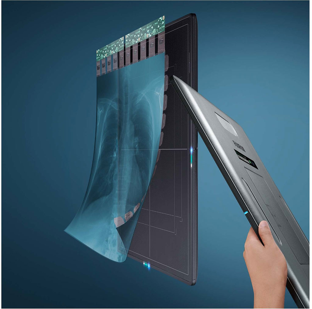
Image: Glass-Free Digital Radiography Detector (Photo courtesy of FUJIFILM Medical Systems U.S.A., Inc.)
FUJIFILM Medical Systems U.S.A., Inc. (Lexington, MA, USA) previewed its new FDR D-EVO III DR detector at the 105th scientific assembly and annual meeting of the Radiological Society of North America (RSNA) held from December 1 – 6, 2019 in Chicago, USA. FUJIFILM Medical Systems, which is part of the FUJIFILM Healthcare portfolio, is a provider of diagnostic imaging products and medical informatics solutions ranging from digital X-ray systems (DR: detectors, mobiles, rooms and 3D mammography), to the comprehensive Synapse Enterprise Imaging portfolio, to full-field digital mammography systems with digital breast tomosynthesis, and computed tomography solutions.
The FDR D-EVO III previewed at RSNA 2019 is FUJIFILM's third-generation digital X-ray detector featuring a sleek, thin design and status as the world's first glass-free DR detector with patented Irradiated Side Sampling (ISS) – also making it the world's lightest 14x17 detector at approximately four pounds. This innovative design removes the traditional glass substrate from the capture layer, eliminating the most fragile layer inside, allowing for a much lighter weight compared to previous models. The FDR D-EVO III 14x17 inch detector incorporates all of the groundbreaking features of the FDR D-EVO II, including its sleek design with smooth and tapered edges for easier positioning, antibacterial nano-coating to help fight against HAIs and a long-lasting battery life.
Additionally, FUJIFILM displayed its full range of DR detectors, including the CALNEO Dual, a 17x17 inch standard cassette sized detector featuring two sensitivity capture layers, coupled with its intelligent energy subtraction processing. A single exposure produces three images; traditional, soft tissue-only, and bone only views. These distinctly different images are expected to be used for visualizing or tracking of lung cancer nodules. The innovative dual capture layer design yields higher definition general X-ray images, enhancing separation accuracy of bone detail and soft tissue.
FUJIFILM also previewed the FDR SE Lite retrofit DR detector at RSNA 2019 that is geared towards bringing DR technology to specialty and small medical practices. Offered as an economical DR solution, the FDR SE Lite DR detector and its companion workstation offer just the right amount of features such as instant image transfer, simplified workflow, and shortened exam times (compared to computed radiography), to meet the unique needs of small practices. Users have the option of upgrading to include Dynamic Visualization II processing which adapts image contrast and density, based on image, thickness and structural recognition.
At RSNA 2019, FUJIFILM also demonstrated its full Synapse portfolio and debut its latest advancement in Enterprise Imaging, Synapse 7x which is a next-generation, secure server-side viewer platform that extends across enterprise imaging areas, bringing diagnostic radiology, mammography and cardiology together through a single, zero footprint platform, and allowing for immediate content interaction regardless of dataset size. Synapse 3D operates natively within the viewer, extending the same advanced visualization across radiology, cardiology, and mammography, eliminating the need for third-party workstations. Synapse 7x is designed to take full advantage of Fujifilm's open AI-enabled platform and use AI results natively within user workflows.
"Fujifilm's Synapse 7x promises to be a game changer for our US healthcare providers. It's a convergence of Fujifilm's server-side technology and was designed to cover all the different areas of diagnostic visualization as well as meet the long-term goal of providing the richest possible visualization layer for an enterprise imaging solution," said Bill Lacy, Vice President of Medical Informatics at FUJIFILM Medical Systems, U.S.A., Inc. "The robust technology is uniquely AI-enabled and integrated, and is presently unrivalled in the marketplace."
Related Links:
FUJIFILM Medical Systems U.S.A., Inc.
Latest RSNA 2019 News
- Carestream Introduces Three-Dimensional Extension of General Radiography Through Its Digital Tomosynthesis Functionality
- Lunit Demonstrates Latest Updated AI Solutions for Chest and Breast Radiology at RSNA 2019
- Bracco Diagnostics Unveils Contrast Media and Device Offerings at RSNA 2019
- Guerbet Showcases New Dose&Care and Other Digital Solutions with Diagnostic and Interventional Imaging Offerings
- Canon Introduces New Wireless Detectors and Digital PET/CT Scanner at RSNA 2019
- Siemens Healthineers Introduces SOMATOM On.site Mobile Head CT Scanner and AI-based MRI Assistants at RSNA
- Hologic Launches Unifi Workspace, Comprehensive Reading Solution for Breast Health Diagnostics
- Agfa Launches New Groundbreaking Digital Radiography Unit at RSNA 2019
- Fujifilm SonoSite Exhibits Complete Point-of-Care Ultrasound Portfolio at RSNA 2019
- NVIDIA Showcases Latest AI-driven Medical Imaging Advancements at RSNA 2019
- Philips Healthcare Demonstrates How AI Breast Software Brings Intelligence and Automation to Breast Ultrasound
- Siemens Healthineers Focuses on Digital Transformation of Imaging and Therapy at RSNA 2019
Channels
Radiography
view channel
Routine Mammograms Could Predict Future Cardiovascular Disease in Women
Mammograms are widely used to screen for breast cancer, but they may also contain overlooked clues about cardiovascular health. Calcium deposits in the arteries of the breast signal stiffening blood vessels,... Read more
AI Detects Early Signs of Aging from Chest X-Rays
Chronological age does not always reflect how fast the body is truly aging, and current biological age tests often rely on DNA-based markers that may miss early organ-level decline. Detecting subtle, age-related... Read moreMRI
view channel
New Material Boosts MRI Image Quality
Magnetic resonance imaging (MRI) is a cornerstone of modern diagnostics, yet certain deep or anatomically complex tissues, including delicate structures of the eye and orbit, remain difficult to visualize clearly.... Read more
AI Model Reads and Diagnoses Brain MRI in Seconds
Brain MRI scans are critical for diagnosing strokes, hemorrhages, and other neurological disorders, but interpreting them can take hours or even days due to growing demand and limited specialist availability.... Read moreMRI Scan Breakthrough to Help Avoid Risky Invasive Tests for Heart Patients
Heart failure patients often require right heart catheterization to assess how severely their heart is struggling to pump blood, a procedure that involves inserting a tube into the heart to measure blood... Read more
MRI Scans Reveal Signature Patterns of Brain Activity to Predict Recovery from TBI
Recovery after traumatic brain injury (TBI) varies widely, with some patients regaining full function while others are left with lasting disabilities. Prognosis is especially difficult to assess in patients... Read moreUltrasound
view channel
Reusable Gel Pad Made from Tamarind Seed Could Transform Ultrasound Examinations
Ultrasound imaging depends on a conductive gel to eliminate air between the probe and the skin so sound waves can pass clearly into the body. While the imaging technology is fast, safe, and noninvasive,... Read more
AI Model Accurately Detects Placenta Accreta in Pregnancy Before Delivery
Placenta accreta spectrum (PAS) is a life-threatening pregnancy complication in which the placenta abnormally attaches to the uterine wall. The condition is a leading cause of maternal mortality and morbidity... Read moreNuclear Medicine
view channel
Radiopharmaceutical Molecule Marker to Improve Choice of Bladder Cancer Therapies
Targeted cancer therapies only work when tumor cells express the specific molecular structures they are designed to attack. In urothelial carcinoma, a common form of bladder cancer, the cell surface protein... Read more
Cancer “Flashlight” Shows Who Can Benefit from Targeted Treatments
Targeted cancer therapies can be highly effective, but only when a patient’s tumor expresses the specific protein the treatment is designed to attack. Determining this usually requires biopsies or advanced... Read moreGeneral/Advanced Imaging
view channel
AI Tool Offers Prognosis for Patients with Head and Neck Cancer
Oropharyngeal cancer is a form of head and neck cancer that can spread through lymph nodes, significantly affecting survival and treatment decisions. Current therapies often involve combinations of surgery,... Read more
New 3D Imaging System Addresses MRI, CT and Ultrasound Limitations
Medical imaging is central to diagnosing and managing injuries, cancer, infections, and chronic diseases, yet existing tools each come with trade-offs. Ultrasound, X-ray, CT, and MRI can be costly, time-consuming,... Read moreImaging IT
view channel
New Google Cloud Medical Imaging Suite Makes Imaging Healthcare Data More Accessible
Medical imaging is a critical tool used to diagnose patients, and there are billions of medical images scanned globally each year. Imaging data accounts for about 90% of all healthcare data1 and, until... Read more
Global AI in Medical Diagnostics Market to Be Driven by Demand for Image Recognition in Radiology
The global artificial intelligence (AI) in medical diagnostics market is expanding with early disease detection being one of its key applications and image recognition becoming a compelling consumer proposition... Read moreIndustry News
view channel
Nuclear Medicine Set for Continued Growth Driven by Demand for Precision Diagnostics
Clinical imaging services face rising demand for precise molecular diagnostics and targeted radiopharmaceutical therapy as cancer and chronic disease rates climb. A new market analysis projects rapid expansion... Read more


