Diffusion-Based MRI May Predict Dementia Advent
|
By MedImaging International staff writers Posted on 24 Oct 2019 |

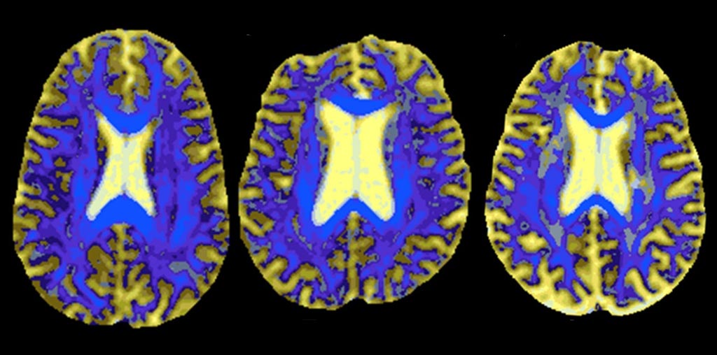
Image: DSEG images of the reference brain (L), a stable SVD patient (C), and a patient who developed dementia (R) (Photo courtesy of Rebecca Charlton/ Goldsmith University of London).
An automatic diffusion tensor image segmentation (DSEG) technique could help assess brain microstructural damage in cerebral small vessel disease (SVD) patients, claims a new study.
Researchers at St George's University of London (SGUL; United Kingdom), Goldsmiths University of London (United Kingdom), and other institutions conducted a study involving 96 SVD patients (aged 43–89 years) in order to explore the extent to which DSEG, which characterizes microstructural damage using just a single diffusion tensor image (DTI) acquisition at 1.5T, can predict both degree of cognitive decline and conversion to dementia. All patients underwent annual MRI scanning for a period of three years and cognitive assessment for a five-year period. DSEG was used to map the cerebrum into 16 segments.
By comparing segments of an individual with SVD to those of a healthy brain, the researchers derived a DSEG spectrum containing information about grey matter, white matter, cerebrospinal fluid (CSF), and regions with diffusion profiles that deviate from those of healthy tissue. They found that DSEG measures increased over time, indicating progression of SVD burden, and that the DSEG measures also predicted decline in executive function and global cognition, as well as identifying stable individuals versus those who developed dementia.
In all, the results revealed that DSEG was significantly related to decline in executive function and global cognition, with 18.2% of the patients converted to dementia. Baseline DSEG predicted dementia with a balanced classification rate of 76%. No relationship was found between DSEG measures and information processing speed; the researchers suggest that perhaps this is because DSEG covers the entire cerebrum and not just white matter tracts, within which information processing and SVD are strongly associated. The study was published on September 12, 2019, in Stroke.
“Our objective was to find a measure of brain tissue microstructural damage. Using a new technique based on readily available MRI scans, we can predict which people go on to show cognitive decline and develop dementia,” said senior author Rebecca Charlton of Goldsmiths University of London. “In the future, DSEG technology could be used as a decision support system for clinicians. This technique has the potential to identify those patients at risk for cognitive decline and vascular dementia.”
Water molecules undergo random Brownian motion, also known as diffusion. MRI is sensitive to this motion, as controlled by the b-value. When the b-value equals zero, the images are not weighted by diffusion; when the b-value is greater than zero the images are diffusion-weighted. When cellular membranes, the myelin shield, etc., hinder the diffusion, the signal is higher. DTI can thus be used to visualize fiber structures, as it can readily differentiate water molecule diffusivities both along and against the fiber.
Related Links:
St George's University of London
Goldsmiths University of London
Latest MRI News
- MRI Scans Reveal Signature Patterns of Brain Activity to Predict Recovery from TBI
- Novel Imaging Approach to Improve Treatment for Spinal Cord Injuries
- AI-Assisted Model Enhances MRI Heart Scans
- AI Model Outperforms Doctors at Identifying Patients Most At-Risk of Cardiac Arrest
- New MRI Technique Reveals Hidden Heart Issues
- Shorter MRI Exam Effectively Detects Cancer in Dense Breasts
- MRI to Replace Painful Spinal Tap for Faster MS Diagnosis
- MRI Scans Can Identify Cardiovascular Disease Ten Years in Advance
- Simple Brain Scan Diagnoses Parkinson's Disease Years Before It Becomes Untreatable
- Cutting-Edge MRI Technology to Revolutionize Diagnosis of Common Heart Problem
- New MRI Technique Reveals True Heart Age to Prevent Attacks and Strokes
- AI Tool Predicts Relapse of Pediatric Brain Cancer from Brain MRI Scans
- AI Tool Tracks Effectiveness of Multiple Sclerosis Treatments Using Brain MRI Scans
- Ultra-Powerful MRI Scans Enable Life-Changing Surgery in Treatment-Resistant Epileptic Patients
- AI-Powered MRI Technology Improves Parkinson’s Diagnoses
- Biparametric MRI Combined with AI Enhances Detection of Clinically Significant Prostate Cancer
Channels
Radiography
view channel
Routine Mammograms Could Predict Future Cardiovascular Disease in Women
Mammograms are widely used to screen for breast cancer, but they may also contain overlooked clues about cardiovascular health. Calcium deposits in the arteries of the breast signal stiffening blood vessels,... Read more
AI Detects Early Signs of Aging from Chest X-Rays
Chronological age does not always reflect how fast the body is truly aging, and current biological age tests often rely on DNA-based markers that may miss early organ-level decline. Detecting subtle, age-related... Read moreUltrasound
view channel
Wearable Ultrasound Imaging System to Enable Real-Time Disease Monitoring
Chronic conditions such as hypertension and heart failure require close monitoring, yet today’s ultrasound imaging is largely confined to hospitals and short, episodic scans. This reactive model limits... Read more
Ultrasound Technique Visualizes Deep Blood Vessels in 3D Without Contrast Agents
Producing clear 3D images of deep blood vessels has long been difficult without relying on contrast agents, CT scans, or MRI. Standard ultrasound typically provides only 2D cross-sections, limiting clinicians’... Read moreNuclear Medicine
view channel
PET Imaging of Inflammation Predicts Recovery and Guides Therapy After Heart Attack
Acute myocardial infarction can trigger lasting heart damage, yet clinicians still lack reliable tools to identify which patients will regain function and which may develop heart failure.... Read more
Radiotheranostic Approach Detects, Kills and Reprograms Aggressive Cancers
Aggressive cancers such as osteosarcoma and glioblastoma often resist standard therapies, thrive in hostile tumor environments, and recur despite surgery, radiation, or chemotherapy. These tumors also... Read more
New Imaging Solution Improves Survival for Patients with Recurring Prostate Cancer
Detecting recurrent prostate cancer remains one of the most difficult challenges in oncology, as standard imaging methods such as bone scans and CT scans often fail to accurately locate small or early-stage tumors.... Read moreGeneral/Advanced Imaging
view channel
AI-Based Tool Accelerates Detection of Kidney Cancer
Diagnosing kidney cancer depends on computed tomography scans, often using contrast agents to reveal abnormalities in kidney structure. Tumors are not always searched for deliberately, as many scans are... Read more
New Algorithm Dramatically Speeds Up Stroke Detection Scans
When patients arrive at emergency rooms with stroke symptoms, clinicians must rapidly determine whether the cause is a blood clot or a brain bleed, as treatment decisions depend on this distinction.... Read moreImaging IT
view channel
New Google Cloud Medical Imaging Suite Makes Imaging Healthcare Data More Accessible
Medical imaging is a critical tool used to diagnose patients, and there are billions of medical images scanned globally each year. Imaging data accounts for about 90% of all healthcare data1 and, until... Read more
Global AI in Medical Diagnostics Market to Be Driven by Demand for Image Recognition in Radiology
The global artificial intelligence (AI) in medical diagnostics market is expanding with early disease detection being one of its key applications and image recognition becoming a compelling consumer proposition... Read moreIndustry News
view channel
GE HealthCare and NVIDIA Collaboration to Reimagine Diagnostic Imaging
GE HealthCare (Chicago, IL, USA) has entered into a collaboration with NVIDIA (Santa Clara, CA, USA), expanding the existing relationship between the two companies to focus on pioneering innovation in... Read more
Patient-Specific 3D-Printed Phantoms Transform CT Imaging
New research has highlighted how anatomically precise, patient-specific 3D-printed phantoms are proving to be scalable, cost-effective, and efficient tools in the development of new CT scan algorithms... Read more
Siemens and Sectra Collaborate on Enhancing Radiology Workflows
Siemens Healthineers (Forchheim, Germany) and Sectra (Linköping, Sweden) have entered into a collaboration aimed at enhancing radiologists' diagnostic capabilities and, in turn, improving patient care... Read more


