Proton Therapy System Available in Multi-Room Configuration
|
By MedImaging International staff writers Posted on 02 Oct 2019 |

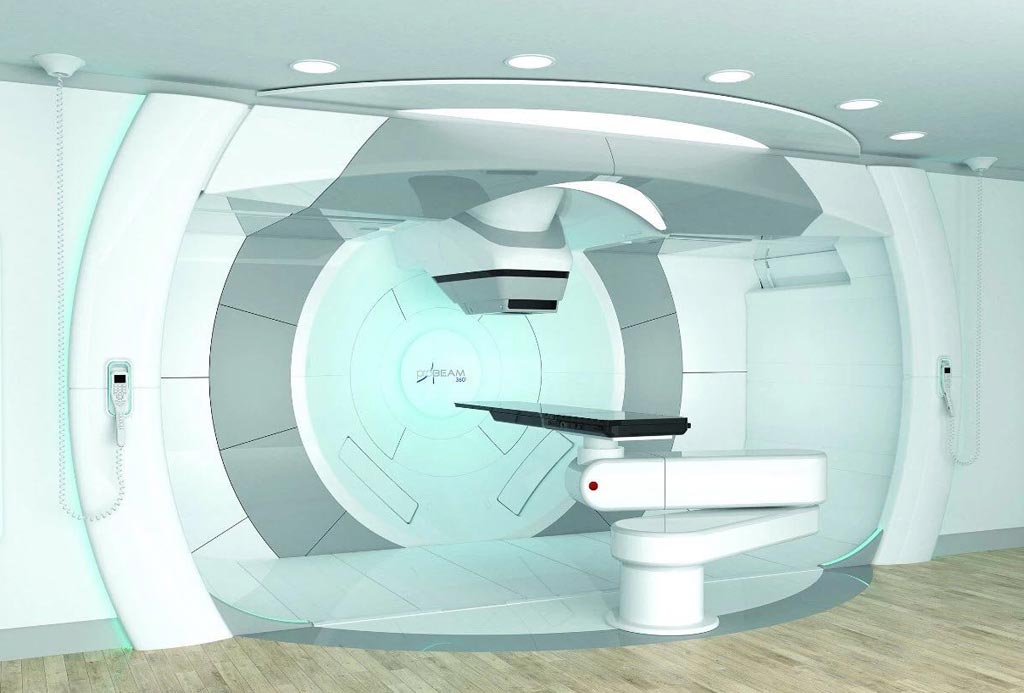
Image: The ProBeam 360° system is now available in multi-room configurations (Photo courtesy of Varian Medical Systems).
The Varian Medical Systems (Varian, Palo Alto, CA, USA) ProBeam 360° system is now available in a multi-room (one to five treatment or research rooms) configuration, which allows cancer centers to tailor the system to meet clinical, research, and capacity needs. Treatment room options include rotating gantries, fixed beam rooms, or eye treatment rooms. Research rooms are available for non-clinical proton beam applications.
The ProBeam 360° system features an extremely powerful cyclotron accelerator, iterative cone-beam computerized tomography (CT) imaging, and high-definition pencil-beam scanning. The 360° rotating gantry enables efficient intensity-modulated proton therapy (IMPT) and faster treatment times by minimizing the need for patient repositioning, thus allowing high-quality cone-beam CT (CBCT) imaging from most angles. RapidScan technology simplifies the process of motion management by delivering each field in a single breath-hold, a capability that increases the number of patients who can comply with breath-hold treatments.
The high-definition pencil-beam scanning technology gives clinicians the ability to deliver dose precisely within the contours of a tumor, minimizing dose to healthy tissue. An iterative CBCT capability enables adaptive precision radiation therapy (RT) during a course of treatment. The system also offers clinicians a viable path to potential next-generation treatments such as Flash therapy. In addition, the ProBeam 360° System offers a 50% smaller volume, a 30% smaller footprint, and 25% lower vault construction cost than Varian’s previous multi-room solution.
“Proton therapy plays an important role in the fight against cancer. The multi-room configuration of ProBeam 360° system provides clinics the flexibility to meet their treatment needs, while also increasing access for patients to proton therapy,” said Kolleen Kennedy, chief growth officer and president of Proton Therapy Solutions at Varian. “Increasing access to advanced treatments like proton therapy is an important step towards achieving our vision of a world without fear of cancer.”
Protons are generated by accelerators in the range of 70 to 250 MeV; by adjusting the energy of the protons during application of treatment, the cell damage due to the proton beam is maximized within the tumor itself. And due to their relatively large mass, protons also have little lateral side scatter in the tissue, staying focused on the tumor. Tissues closer to the surface of the body than the tumor receive reduced radiation, while tissues deeper within the body receive very few protons, so that the dosage becomes immeasurably small.
Latest Nuclear Medicine News
- PET Imaging of Inflammation Predicts Recovery and Guides Therapy After Heart Attack
- Radiotheranostic Approach Detects, Kills and Reprograms Aggressive Cancers
- New Imaging Solution Improves Survival for Patients with Recurring Prostate Cancer
- PET Tracer Enables Same-Day Imaging of Triple-Negative Breast and Urothelial Cancers
- New Camera Sees Inside Human Body for Enhanced Scanning and Diagnosis
- Novel Bacteria-Specific PET Imaging Approach Detects Hard-To-Diagnose Lung Infections
- New Imaging Approach Could Reduce Need for Biopsies to Monitor Prostate Cancer
- Novel Radiolabeled Antibody Improves Diagnosis and Treatment of Solid Tumors
- Novel PET Imaging Approach Offers Never-Before-Seen View of Neuroinflammation
- Novel Radiotracer Identifies Biomarker for Triple-Negative Breast Cancer
- Innovative PET Imaging Technique to Help Diagnose Neurodegeneration
- New Molecular Imaging Test to Improve Lung Cancer Diagnosis
- Novel PET Technique Visualizes Spinal Cord Injuries to Predict Recovery
- Next-Gen Tau Radiotracers Outperform FDA-Approved Imaging Agents in Detecting Alzheimer’s
- Breakthrough Method Detects Inflammation in Body Using PET Imaging
- Advanced Imaging Reveals Hidden Metastases in High-Risk Prostate Cancer Patients
Channels
Radiography
view channel
Routine Mammograms Could Predict Future Cardiovascular Disease in Women
Mammograms are widely used to screen for breast cancer, but they may also contain overlooked clues about cardiovascular health. Calcium deposits in the arteries of the breast signal stiffening blood vessels,... Read more
AI Detects Early Signs of Aging from Chest X-Rays
Chronological age does not always reflect how fast the body is truly aging, and current biological age tests often rely on DNA-based markers that may miss early organ-level decline. Detecting subtle, age-related... Read moreMRI
view channel
Novel Imaging Approach to Improve Treatment for Spinal Cord Injuries
Vascular dysfunction in the spinal cord contributes to multiple neurological conditions, including traumatic injuries and degenerative cervical myelopathy, where reduced blood flow can lead to progressive... Read more
AI-Assisted Model Enhances MRI Heart Scans
A cardiac MRI can reveal critical information about the heart’s function and any abnormalities, but traditional scans take 30 to 90 minutes and often suffer from poor image quality due to patient movement.... Read more
AI Model Outperforms Doctors at Identifying Patients Most At-Risk of Cardiac Arrest
Hypertrophic cardiomyopathy is one of the most common inherited heart conditions and a leading cause of sudden cardiac death in young individuals and athletes. While many patients live normal lives, some... Read moreUltrasound
view channel
Wearable Ultrasound Imaging System to Enable Real-Time Disease Monitoring
Chronic conditions such as hypertension and heart failure require close monitoring, yet today’s ultrasound imaging is largely confined to hospitals and short, episodic scans. This reactive model limits... Read more
Ultrasound Technique Visualizes Deep Blood Vessels in 3D Without Contrast Agents
Producing clear 3D images of deep blood vessels has long been difficult without relying on contrast agents, CT scans, or MRI. Standard ultrasound typically provides only 2D cross-sections, limiting clinicians’... Read moreGeneral/Advanced Imaging
view channel
AI-Based Tool Accelerates Detection of Kidney Cancer
Diagnosing kidney cancer depends on computed tomography scans, often using contrast agents to reveal abnormalities in kidney structure. Tumors are not always searched for deliberately, as many scans are... Read more
New Algorithm Dramatically Speeds Up Stroke Detection Scans
When patients arrive at emergency rooms with stroke symptoms, clinicians must rapidly determine whether the cause is a blood clot or a brain bleed, as treatment decisions depend on this distinction.... Read moreImaging IT
view channel
New Google Cloud Medical Imaging Suite Makes Imaging Healthcare Data More Accessible
Medical imaging is a critical tool used to diagnose patients, and there are billions of medical images scanned globally each year. Imaging data accounts for about 90% of all healthcare data1 and, until... Read more
Global AI in Medical Diagnostics Market to Be Driven by Demand for Image Recognition in Radiology
The global artificial intelligence (AI) in medical diagnostics market is expanding with early disease detection being one of its key applications and image recognition becoming a compelling consumer proposition... Read moreIndustry News
view channel
GE HealthCare and NVIDIA Collaboration to Reimagine Diagnostic Imaging
GE HealthCare (Chicago, IL, USA) has entered into a collaboration with NVIDIA (Santa Clara, CA, USA), expanding the existing relationship between the two companies to focus on pioneering innovation in... Read more
Patient-Specific 3D-Printed Phantoms Transform CT Imaging
New research has highlighted how anatomically precise, patient-specific 3D-printed phantoms are proving to be scalable, cost-effective, and efficient tools in the development of new CT scan algorithms... Read more
Siemens and Sectra Collaborate on Enhancing Radiology Workflows
Siemens Healthineers (Forchheim, Germany) and Sectra (Linköping, Sweden) have entered into a collaboration aimed at enhancing radiologists' diagnostic capabilities and, in turn, improving patient care... Read more


