GE Healthcare Partners with Fujitsu to Develop AI for Spotting Brain Aneurysms
|
By MedImaging International staff writers Posted on 22 Aug 2019 |


|
GE Healthcare (Chicago, IL, USA) has entered into a research collaboration with Fujitsu Australia (Tokyo, Japan) to diagnose and monitor brain aneurysms on scans faster and more efficiently by using artificial intelligence (AI). The project has already received a Cooperative Research Centres Projects (CRC-P) grant of AUD 2.1 million from the Department of Industry, Innovation and Science.
Fujitsu will lead the initiative and leverage its AI and digital solutions capability through its dedicated innovation team in Australia. GE Healthcare will contribute through its leading medical imaging technology. Fujitsu will apply AI methods to images of the brain generated by GE’s Revolution CT scanner, and use a specifically trained algorithm to look for abnormalities and aneurysms. Macquarie University and Macquarie Medical Imaging will provide clinical expertise for the development and testing of the technology. Initially the project will focus on refining the technology with a view to creating a fully commercialized solution that will be distributed initially through radiology practices in Australia and eventually on a worldwide basis.
The outcomes from this project are anticipated to include the development and validation of an AI algorithm capable of highlighting blood vessels within the circle of Willis, an arterial ring sited at the base of the brain that may have one or more aneurysms. This technology will also allow the tracking of identified aneurysms over time, providing radiologists with a valuable diagnostic support tool and patients with greater peace of mind that known aneurysms are being effectively monitored over the long-term. A second element to the project will include a planning tool for surgical (stent) intervention. This tool will use fluid dynamic modeling to predict the risk of aneurysm rupture.
“We are pleased to be part of this important ‘co-creation’ initiative that leverages the strengths of each of our partners, as well as Fujitsu’s experience in AI to have a positive impact on peoples’ lives,” said Mike Foster, Chief Executive Officer of Fujitsu Australia and New Zealand. “AI in particular has the capability to make our daily lives more comfortable and contribute to solving difficult problems such as detecting serious medical issues early and allowing more timely treatment intervention. This is an excellent demonstration of Fujitsu’s commitment to creating human-centric innovation together with our customers and partners to build a trusted future where everyone can feel safe.”
“As the consequences of brain aneurysm rupture are often fatal, effective and expedient detection is crucial. Unfortunately screening and monitoring takes time and specialist expertise not afforded by every radiology practice,” said Matt Tucker, President and CEO GE Healthcare Australia & New Zealand. “The application of AI can give doctors better insights more quickly and produce fewer variable results. We are proud to be part of this program that will transform brain aneurysm diagnosis and improve patient outcomes.”
“This is an excellent example of the MQ Health model of “Heal, Learn and Discover” in action with industry. Macquarie University, with its own hospital and clinical expertise is well placed to actively contribute to the development of applied medical innovations and welcomes the opportunity to work with leading information technology, healthcare and diagnostic companies such as Fujitsu, GE Healthcare and Macquarie Medical Imaging,” said Professor Patrick McNeil, Deputy Vice Chancellor Medicine and Health and Executive Dean, Faculty of Medicine and Health Sciences, Macquarie University.


 Guided Devices.jpg)



Fujitsu will lead the initiative and leverage its AI and digital solutions capability through its dedicated innovation team in Australia. GE Healthcare will contribute through its leading medical imaging technology. Fujitsu will apply AI methods to images of the brain generated by GE’s Revolution CT scanner, and use a specifically trained algorithm to look for abnormalities and aneurysms. Macquarie University and Macquarie Medical Imaging will provide clinical expertise for the development and testing of the technology. Initially the project will focus on refining the technology with a view to creating a fully commercialized solution that will be distributed initially through radiology practices in Australia and eventually on a worldwide basis.
The outcomes from this project are anticipated to include the development and validation of an AI algorithm capable of highlighting blood vessels within the circle of Willis, an arterial ring sited at the base of the brain that may have one or more aneurysms. This technology will also allow the tracking of identified aneurysms over time, providing radiologists with a valuable diagnostic support tool and patients with greater peace of mind that known aneurysms are being effectively monitored over the long-term. A second element to the project will include a planning tool for surgical (stent) intervention. This tool will use fluid dynamic modeling to predict the risk of aneurysm rupture.
“We are pleased to be part of this important ‘co-creation’ initiative that leverages the strengths of each of our partners, as well as Fujitsu’s experience in AI to have a positive impact on peoples’ lives,” said Mike Foster, Chief Executive Officer of Fujitsu Australia and New Zealand. “AI in particular has the capability to make our daily lives more comfortable and contribute to solving difficult problems such as detecting serious medical issues early and allowing more timely treatment intervention. This is an excellent demonstration of Fujitsu’s commitment to creating human-centric innovation together with our customers and partners to build a trusted future where everyone can feel safe.”
“As the consequences of brain aneurysm rupture are often fatal, effective and expedient detection is crucial. Unfortunately screening and monitoring takes time and specialist expertise not afforded by every radiology practice,” said Matt Tucker, President and CEO GE Healthcare Australia & New Zealand. “The application of AI can give doctors better insights more quickly and produce fewer variable results. We are proud to be part of this program that will transform brain aneurysm diagnosis and improve patient outcomes.”
“This is an excellent example of the MQ Health model of “Heal, Learn and Discover” in action with industry. Macquarie University, with its own hospital and clinical expertise is well placed to actively contribute to the development of applied medical innovations and welcomes the opportunity to work with leading information technology, healthcare and diagnostic companies such as Fujitsu, GE Healthcare and Macquarie Medical Imaging,” said Professor Patrick McNeil, Deputy Vice Chancellor Medicine and Health and Executive Dean, Faculty of Medicine and Health Sciences, Macquarie University.

MRI System
nanoScan MRI 3T/7T
Ultrasound-Guided Biopsy & Visualization Tools
Endoscopic Ultrasound (EUS) Guided Devices
Diagnostic Ultrasound System
DC-80A
Digital Color Doppler Ultrasound System
MS22Plus
Latest Industry News News
- GE HealthCare Showcases AI-Enabled Nuclear Medicine Portfolio at SNMMI 2026
- GE HealthCare Highlights AI-Supported Radiation Therapy Tools at ESTRO 2026
- Nuclear Medicine Set for Continued Growth Driven by Demand for Precision Diagnostics
- GE HealthCare and NVIDIA Collaboration to Reimagine Diagnostic Imaging
- Patient-Specific 3D-Printed Phantoms Transform CT Imaging
- Siemens and Sectra Collaborate on Enhancing Radiology Workflows
- Bracco Diagnostics and ColoWatch Partner to Expand Availability CRC Screening Tests Using Virtual Colonoscopy
- Mindray Partners with TeleRay to Streamline Ultrasound Delivery
- Philips and Medtronic Partner on Stroke Care
- Siemens and Medtronic Enter into Global Partnership for Advancing Spine Care Imaging Technologies
- RSNA 2024 Technical Exhibits to Showcase Latest Advances in Radiology
- Bracco Collaborates with Arrayus on Microbubble-Assisted Focused Ultrasound Therapy for Pancreatic Cancer
- Innovative Collaboration to Enhance Ischemic Stroke Detection and Elevate Standards in Diagnostic Imaging
- RSNA 2024 Registration Opens
- Microsoft collaborates with Leading Academic Medical Systems to Advance AI in Medical Imaging
- GE HealthCare Acquires Intelligent Ultrasound Group’s Clinical Artificial Intelligence Business
Channels
Radiography
view channel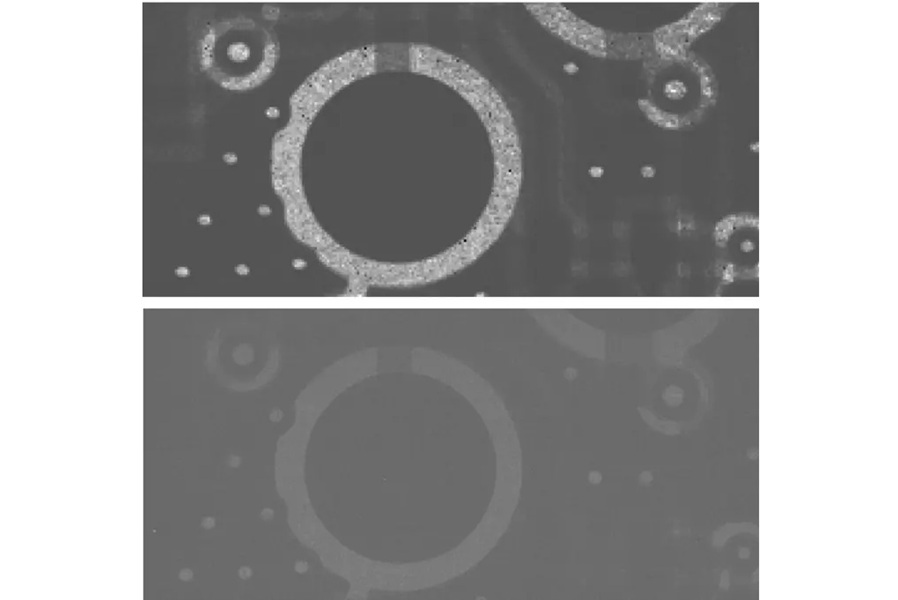
Computational X-Ray Technique Enables High-Resolution, Low-Dose Radiography
Reducing radiation in medical X-ray imaging remains a persistent challenge because dose reductions often degrade spatial resolution and diagnostic confidence. This is especially critical in pediatrics,... Read more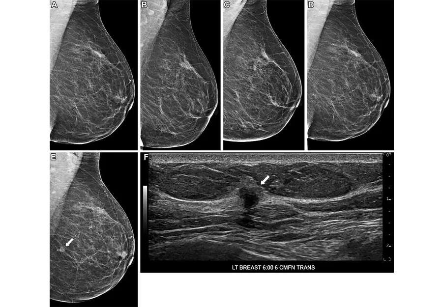
AI Tool Predicts Five-Year Breast Cancer Risk from Mammograms
Breast cancer risk assessment during routine screening is difficult because many women who develop the disease have no known genetic mutations or family history. Static risk tools provide limited discrimination... Read more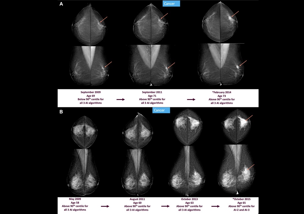
MRI
view channel
Advanced MRI Reveals Structural Brain Changes Linked to Better Cognitive Health
Objective, noninvasive measures that capture subtle, training-related changes in adult brain structure remain limited in routine clinical practice. Quantifying neuroplasticity with precision is especially... Read more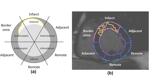
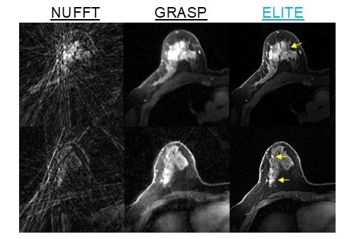
Cardiac MRI Detects Hidden Heart Dysfunction After Heart Attack
Myocardial infarction often leaves patients with persistent ventricular dysfunction that can progress without obvious signs. Detecting early decline remains difficult because standard measures may miss... Read more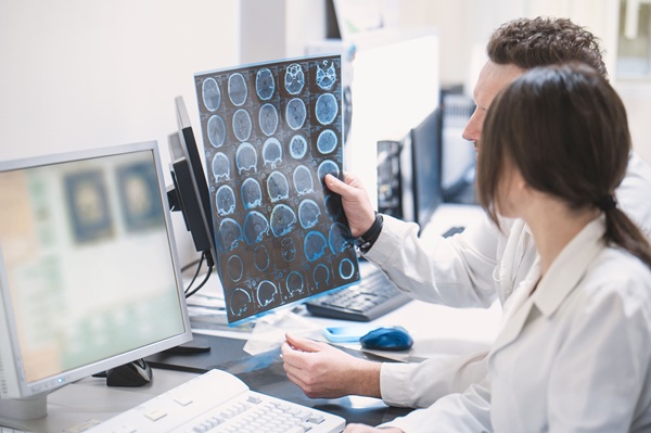
Ultrasound
view channel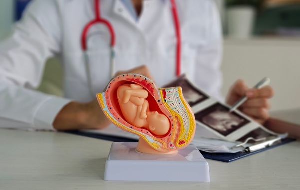
Simple 2D Ultrasound Tool Accurately Estimates Placental Volume
Stillbirth remains a major cause of perinatal loss, with about 21,000 cases annually in the United States. Although placental abnormalities are linked to adverse pregnancy outcomes, routine monitoring... Read more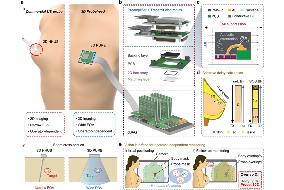
Portable 3D Ultrasound System Enables Reproducible Breast Cancer Monitoring
Breast cancer can develop between annual mammograms, and these interval cancers account for 20% to 30% of cases and tend to be more aggressive. The challenge is pronounced in people with dense breast tissue.... Read more
Nuclear Medicine
view channel
Radiotherapy Professionals Embrace AI as a Clinical Support Tool
Radiotherapy planning requires precise identification of organs at risk near a tumor, but manual contouring is repetitive and time-consuming, often delaying plan completion and straining specialist capacity.... Read more
Second PSMA PET Identifies Disease in More Than Half of Negative Cases
Biochemical recurrence of prostate cancer after prostatectomy or radiation can be difficult to localize when imaging is negative. Uncertain disease sites may delay salvage therapy and expose patients to... Read more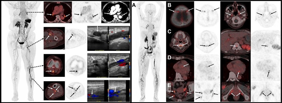
![Image: (A-C) Representative whole-body maximum-intensity projection images and regional fused PET/CT images from three histologically confirmed osteosarcoma patients who underwent paired [68Ga]Ga-B7-H3-BCH PET/CT and 18F-FDGE PET/CT within 7 days. (D) Multimodal imaging evaluation of patient 3, including X-ray, MRI (T2-weighted imaging, T2WI), CT, and B7-H3 PET/CT. (Photo courtesy of SNMMI)](https://globetechcdn.com/mobile_medicalimaging/images/stories/articles/article_images/2026-06-07/294811110_image_1.jpg "Image: (A-C) Representative whole-body maximum-intensity projection images and regional fused PET/CT images from three histologically confirmed osteosarcoma patients who underwent paired [68Ga]Ga-B7-H3-BCH PET/CT and 18F-FDGE PET/CT within 7 days. (D) Multimodal imaging evaluation of patient 3, including X-ray, MRI (T2-weighted imaging, T2WI), CT, and B7-H3 PET/CT. (Photo courtesy of SNMMI)")
General/Advanced Imaging
view channel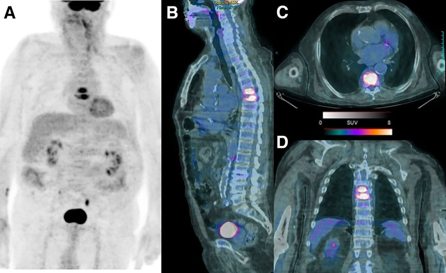
PET/CT Improves Diagnosis and Management of Fever of Unknown Origin
Fever of unknown origin (FUO) is persistent fever without an identifiable cause and remains difficult to diagnose because potential etiologies are broad and no single gold-standard pathway exists.... Read more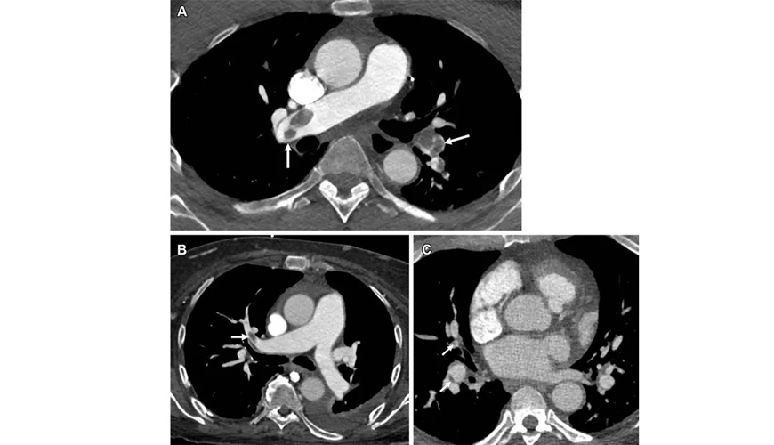
New PE-RADS Framework Standardizes Pulmonary Embolism Imaging Reports
Pulmonary embolism is a sudden blockage of the lung’s blood vessels that reduces pulmonary blood flow, can strain the right hear,t and may be fatal if untreated. Patients often present with shortness of... Read more
Imaging IT
view channel
Ambient AI Reporting Platform Streamlines Radiology Reporting
Radiology departments face growing imaging volumes and staffing shortages, creating reporting bottlenecks and pressure to maintain turnaround times. Conventional dictation tools document findings after... Read more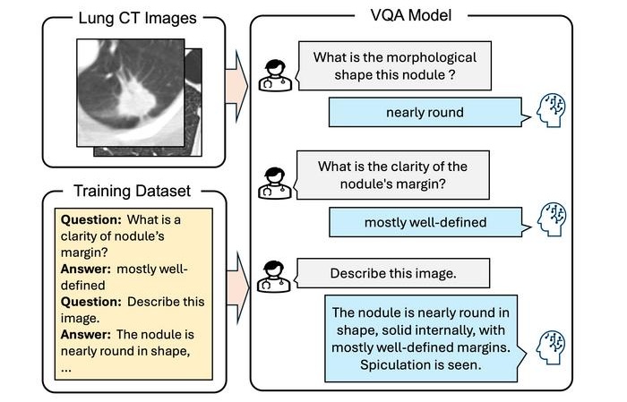
Interactive AI Tool Supports Explainable Lung Nodule Assessment
Lung cancer is a leading cause of cancer mortality, and timely characterization of pulmonary nodules on chest computed tomography (CT) is essential for directing care. Interpreting nodule morphology demands... Read more