Konica Minolta Offers AI-Based Cardiac Ultrasound Analysis
|
By MedImaging International staff writers Posted on 06 Jul 2019 |

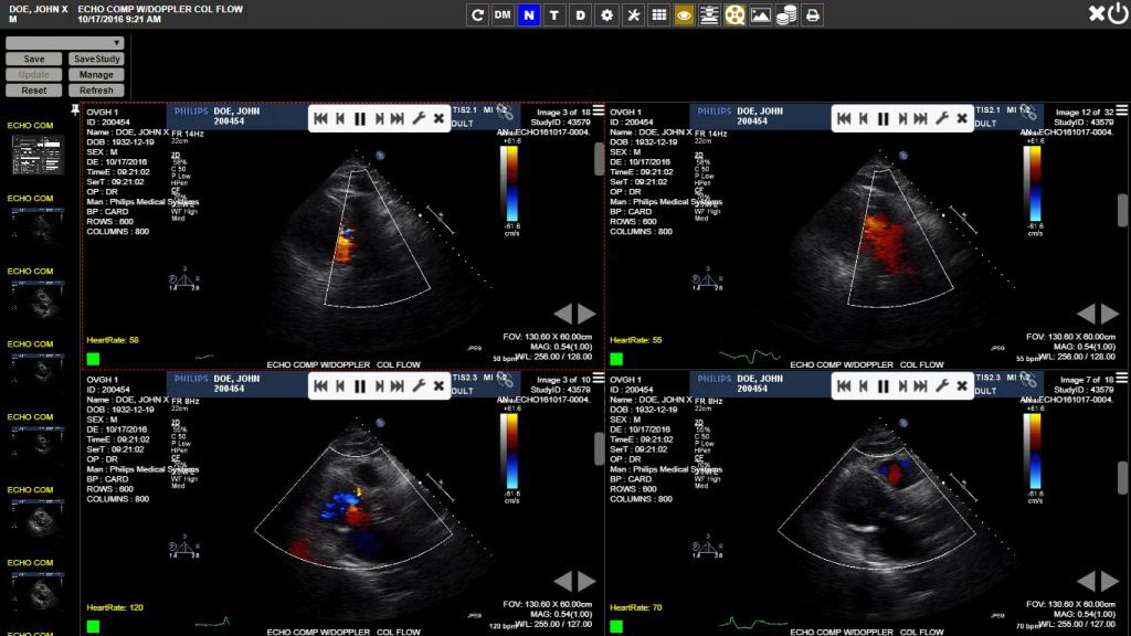
Image: A screenshot from the Exa Cardio PACS Platform (Photo courtesy of Konica Minolta Healthcare).
Konica Minolta Healthcare Americas Inc, (Wayne, NJ, USA), a provider of medical diagnostic imaging and healthcare information technology, has announced a partnership with DiA Imaging Analysis (BE'ER SHEVA, Israel), a provider of artificial intelligence (AI) powered ultrasound analysis solutions. The partnership aims to expand the analysis capabilities of Konica Minolta's Exa Cardio PACS Platform (Cardiovascular Information System) with DiA's cardiac analysis, "LVivo Toolbox."
DiA's LVivo Cardiac Toolbox uses AI-based technology to analyze cardiac ultrasound images automatically and objectively, to reduce the subjectivity of manual or visual analysis methods used today. The LVivo Cardiac Toolbox uses novel pattern recognition, deep-learning and machine-learning algorithms that automatically imitate how the human eye detects borders and motion. DiA's LVivo Cardiac Toolbox is vendor-neutral, supporting DICOM clips of various ultrasound systems.
Konica Minolta's Exa platform provides hospitals and imaging centers with the ability to view DICOM and non-DICOM images and information across departments and facilities, regardless of the original source. Exa also provides a vendor-neutral centralized archive and image exchange. Konica Minolta will offer the LVivo Toolbox as a part of Exa's diagnostic-quality Zero Footprint, Server Side Rendering Universal Viewer for DICOM and non-DICOM images. The integration has been designed according to Exa's user interface to assure efficient workflow and accessibility to all Exa Cardio PACS users.
"With DiA's LVivo Toolbox, Konica Minolta offers clinicians decision support with objective data," said Andrew Horning, Konica Minolta's Cardiology Product Manager. "Through this partnership, we integrate innovative, AI-based cardiac analysis into Exa's already powerful and user-customizable structured reporting system; all available anywhere - from a multi-monitor workstation on a hospital network, to a laptop PC on Wi-Fi. This helps cardiologists make better decisions sooner."
"We are excited to partner with Konica and offer the LVivo Cardiac Toolbox to Exa Cardio PACS users," said Hila Goldman Aslan, DiA's CEO and Co-Founder. "Konica Minolta is a world leader in providing innovative, state-of-the-art healthcare solutions to its customers. With LVivo Toolbox offered as a part of Exa Cardio PACS, Konica's users will be able to get fast, valuable and objective insights into cardiac function analysis. Our mission is to support clinicians with various cardiac analysis experience levels, in their decision-making process and to use our solutions as part of their everyday analysis routine."
Latest Industry News News
- Nuclear Medicine Set for Continued Growth Driven by Demand for Precision Diagnostics
- GE HealthCare and NVIDIA Collaboration to Reimagine Diagnostic Imaging
- Patient-Specific 3D-Printed Phantoms Transform CT Imaging
- Siemens and Sectra Collaborate on Enhancing Radiology Workflows
- Bracco Diagnostics and ColoWatch Partner to Expand Availability CRC Screening Tests Using Virtual Colonoscopy
- Mindray Partners with TeleRay to Streamline Ultrasound Delivery
- Philips and Medtronic Partner on Stroke Care
- Siemens and Medtronic Enter into Global Partnership for Advancing Spine Care Imaging Technologies
- RSNA 2024 Technical Exhibits to Showcase Latest Advances in Radiology
- Bracco Collaborates with Arrayus on Microbubble-Assisted Focused Ultrasound Therapy for Pancreatic Cancer
- Innovative Collaboration to Enhance Ischemic Stroke Detection and Elevate Standards in Diagnostic Imaging
- RSNA 2024 Registration Opens
- Microsoft collaborates with Leading Academic Medical Systems to Advance AI in Medical Imaging
- GE HealthCare Acquires Intelligent Ultrasound Group’s Clinical Artificial Intelligence Business
- Bayer and Rad AI Collaborate on Expanding Use of Cutting Edge AI Radiology Operational Solutions
- Polish Med-Tech Company BrainScan to Expand Extensively into Foreign Markets
Channels
Radiography
view channel
Routine Mammograms Could Predict Future Cardiovascular Disease in Women
Mammograms are widely used to screen for breast cancer, but they may also contain overlooked clues about cardiovascular health. Calcium deposits in the arteries of the breast signal stiffening blood vessels,... Read more
AI Detects Early Signs of Aging from Chest X-Rays
Chronological age does not always reflect how fast the body is truly aging, and current biological age tests often rely on DNA-based markers that may miss early organ-level decline. Detecting subtle, age-related... Read moreMRI
view channel
MRI Scans Reveal Signature Patterns of Brain Activity to Predict Recovery from TBI
Recovery after traumatic brain injury (TBI) varies widely, with some patients regaining full function while others are left with lasting disabilities. Prognosis is especially difficult to assess in patients... Read more
Novel Imaging Approach to Improve Treatment for Spinal Cord Injuries
Vascular dysfunction in the spinal cord contributes to multiple neurological conditions, including traumatic injuries and degenerative cervical myelopathy, where reduced blood flow can lead to progressive... Read more
AI-Assisted Model Enhances MRI Heart Scans
A cardiac MRI can reveal critical information about the heart’s function and any abnormalities, but traditional scans take 30 to 90 minutes and often suffer from poor image quality due to patient movement.... Read more
AI Model Outperforms Doctors at Identifying Patients Most At-Risk of Cardiac Arrest
Hypertrophic cardiomyopathy is one of the most common inherited heart conditions and a leading cause of sudden cardiac death in young individuals and athletes. While many patients live normal lives, some... Read moreUltrasound
view channel
Portable Ultrasound Sensor to Enable Earlier Breast Cancer Detection
Breast cancer screening relies heavily on annual mammograms, but aggressive tumors can develop between scans, accounting for up to 30 percent of cases. These interval cancers are often diagnosed later,... Read more
Portable Imaging Scanner to Diagnose Lymphatic Disease in Real Time
Lymphatic disorders affect hundreds of millions of people worldwide and are linked to conditions ranging from limb swelling and organ dysfunction to birth defects and cancer-related complications.... Read more
Imaging Technique Generates Simultaneous 3D Color Images of Soft-Tissue Structure and Vasculature
Medical imaging tools often force clinicians to choose between speed, structural detail, and functional insight. Ultrasound is fast and affordable but typically limited to two-dimensional anatomy, while... Read moreNuclear Medicine
view channel
Radiopharmaceutical Molecule Marker to Improve Choice of Bladder Cancer Therapies
Targeted cancer therapies only work when tumor cells express the specific molecular structures they are designed to attack. In urothelial carcinoma, a common form of bladder cancer, the cell surface protein... Read more
Cancer “Flashlight” Shows Who Can Benefit from Targeted Treatments
Targeted cancer therapies can be highly effective, but only when a patient’s tumor expresses the specific protein the treatment is designed to attack. Determining this usually requires biopsies or advanced... Read moreGeneral/Advanced Imaging
view channel
AI Tool Offers Prognosis for Patients with Head and Neck Cancer
Oropharyngeal cancer is a form of head and neck cancer that can spread through lymph nodes, significantly affecting survival and treatment decisions. Current therapies often involve combinations of surgery,... Read more
New 3D Imaging System Addresses MRI, CT and Ultrasound Limitations
Medical imaging is central to diagnosing and managing injuries, cancer, infections, and chronic diseases, yet existing tools each come with trade-offs. Ultrasound, X-ray, CT, and MRI can be costly, time-consuming,... Read moreImaging IT
view channel
New Google Cloud Medical Imaging Suite Makes Imaging Healthcare Data More Accessible
Medical imaging is a critical tool used to diagnose patients, and there are billions of medical images scanned globally each year. Imaging data accounts for about 90% of all healthcare data1 and, until... Read more


